Intranasal delivery of SARS-CoV-2 S RDB protein causes severe histological damage in the adult zebrafish OO that recovers after 2 weeks. H&E stains of zebrafish OO paraffin sections. (A) Control vehicle treated OO. (B) 3 h; (C) 1 d; (D) 3 d; (E) 5 d; (F) 2 w post intranasal delivery of SARS-CoV-2 S RDB protein (r-Spike) or controls (Poly(I:C) and an off-target His-tagged recombinant protein). Images are representative of n=10 sections per animal and N=3-5 fish per experimental group. Asterisk denotes edemic spaces in the olfactory lamellae, mostly in the lamina propria. He: hemorrhages. Arrow heads: apical loss of olfactory sensory neurons. Black Arrow: Necrotic lamella. (G) Immunofluorescence image of an AB zebrafish OO cryosection from an adult OMP:lynRFP animal 15 min after I.N SARS-CoV-2 S RBD administration stained with a rabbit IgG isotype control as primary antibody. Scale: 20μm. (H) Immunofluorescence image of an OMP:lynRFP zebrafish OO stained with anti-His antibody (green) 15 min post I.N. delivery of recombinant His-tagged SARS-CoV-2 S RBD. (I) Immunofluorescence of an AB zebrafish OO stained with anti-His antibody (red) 15 min post I.N. delivery of SARS-CoV-2 S RBD shows puncta of anti-His staining when zoomed in on non-sensory epithelium; Scale = 5μm. Ax: OSN axon bundles; NC: nasal cavity; LP: lamina propria. Scale: 20μm. Cell nuclei are stained with DAPI (blue). (J) Mean width of the olfactory lamina propria in each treatment group. (K) Mean percent of olfactory lamellar surface covered by healthy cilia in each treatment group. (L) Expression of ace2 in OO and OB tissue of untreated adult AB zebrafish (=3) as measured by RT-qPCR. Expression levels were normalized to rps11 as the house-keeping gene. Quantifications in J&K were performed in ImageJ; error bars represent S.E.M. J&K statistics were done by one-way ANOVA followed by a Tukey’s post hoc comparison. * P-value<0.05, ** P-value<0.01, **** P-value<0.0001.

Intranasal delivery of SARS-CoV-2 S RDB protein causes severe cellular changes to the OO of adult zebrafish. (A) H&E image of raphe of the OO showing large eosinophilic deposits 3 d post intranasal delivery of SARS-CoV-2 S RBD (B) Masson Trichrome stains of zebrafish OO administered intranasally vehicle or (C-D) 3 h SARS-CoV-2 S RBD, or (E-F) 3 d SARS-CoV-2 S RBD. Asterisk denotes red keratin deposits. Blue staining corresponds to collagen deposits. (G) anti-CTK8 (red) immunofluorescence staining of adult zebrafish OO cryosections after intranasal delivery of vehicle or (H) 3 h; or (I) 3 d SARS-CoV-2 S RBD. (J) anti-PCNA immunofluorescence staining of adult zebrafish OO cryosection after intranasal delivery of vehicle or (K) 3 h; or (L) 3 d recombinant SARS-CoV-2 S RBD. (M) anti-caspase3 (red) immunofluorescence staining of adult zebrafish OO cryosections after intranasal delivery of vehicle or (N) 3 h; (O) 3 d recombinant SARS-CoV-2 S RBD. Nuclei are stained with DAPI (blue). Scale in (G-O): 20μm. (P) Quantification of the mean number of caspase3 positive cells per field of view. (Q) Quantification of number of PCNA positive cells per field of view. Quantifications shown in P and Q were performed on N = 4 animals and n=10 fields of view per animal, represented by different colors. Error bars represent S.E.M. LP: lamina propria; NC: nasal cavity. Statistics ANOVA followed by a Tukey’s post hoc comparison. *P-value<0.05. Representative images of Poly(I:C)-treated animals are shown in Supplemental figure 2.

Intranasal delivery of SARS-CoV-2 S RDB protein causes anosmia in adult zebrafish. (A) Representative EOG of adult zebrafish showing olfactory responses to food over time before and after addition of SARS-CoV-2 S RDB protein (yellow) or a control recombinant protein (black) to the naris normalized to pre-exposure values. (B) Mean percentage loss of olfactory detection of food or bile salts measured by EOG recording. Adult zebrafish (N=4) were treated with SARS-CoV-2 S RDB protein in one naris or vehicle control (PBS) in the other naris for 3 h (yellow), 1 d (teal), 3 d (purple) or 2 w (green) after which EOG were recorded by exposing animals to either food odorant or bile and recording electrical responses in each individual naris. The percent reduction was calculated as percent of the SARS-CoV-2 S RDB treated naris EOG response to vehicle treated naris EOG response. (C) Mean percentage loss of olfactory detection of food or bile salts measured by EOG recording. Adult zebrafish (N=4) were treated with control recombinant protein (r-CK12a) in one naris or vehicle control (PBS) in the other naris for 3 h, 1 d, 3 d or 2 w after which EOG were recorded by exposing animals to either food or bile and recording electrical responses in each individual naris. The percent reduction was calculated as percent of the control recombinant treated naris EOG response to vehicle treated naris EOG response. (D) Graphical representation of the behavioral choice test used in this study. (E) Calculated average difference in time spent on the half of arena containing food odorant or on the half of the arena containing vehicle odorant after adult zebrafish were treated I.N. with vehicle, 3 h or 3 d SARS-CoV-2, or 3 h or 3 d control recombinant (r-CK12a). Red dotted line represents average difference if fish displays zero preference for either odor. Statistical analyses for (B & C) were determined by ANOVA with Tukey’s post hoc multiple comparison whereas a Wilcoxon match pairs signed-rank test was used for behavioral analysis in (E). Error bars in B & C represent S.E.M. and error bars in E represent 95% confidence interval (CI). * P-value <0.05; ** P-value <0.01; *** P-value < 0.001.

ScRNA-Seq of zebrafish OO reveals changes in cellular landscape due to intranasal SARS-CoV-2 S RBD protein delivery. (A-B) Cellular composition of the zebrafish OO visualized using t-distributed stochastic neighbor embedding (tSNE) of vehicle treated, 3 h and 3 d post-intranasal delivery of SARS-CoV-2 S RBD (r-Spike). Individual single-cell transcriptomes were colored according to cluster identity. tSNE plots were generated in Seurat and identified 20 and 22 distinct cell clusters, respectively. (C) tSNE plots of zebrafish OO cell suspensions colored according to treatment. (D) Bar plots showing the proportion of cells belonging to each cluster in each treatment group. Statistical analysis for changes in cell population proportions is in supplemental tables 3 and 4. (E-F) Violin plots showing expression of selected markers of inflammation and vasculature remodeling in the 3 h SARS-CoV-2 S RBD (orange) versus vehicle treated (blue) and 3 d SARS-CoV-2 S RBD (green) versus vehicle treated (blue) zebrafish OO obtained by scRNA-Seq. All represented genes were significantly differentially regulated between treatments (adjusted P-value-adj <0.05).

Intranasal SARS-CoV-2 S RBD protein delivery causes widespread losses of olfactory receptor expression in adult zebrafish. (A-B) Bar plots showing fold-change in expression of olfactory receptor genes in the zebrafish OO 3 h and 3 d post intranasal SARS-CoV-2 S RBD protein compared to vehicle controls identified by scRNA-Seq. (C-D) Violin plots showing changes in significantly differentially expressed ompb expression, a marker of mature ciliated OSNs, in zebrafish OO cell suspensions, across all olfactory neuronal cell clusters in 3 h SARS-CoV-2 S RBD (orange) and 3 d SARS-CoV-2 S RBD (green) versus vehicle treated (blue). (E-F) Summary webs of gene ontology analyses performed in ShinyGO v0.61 from the scRNA-Seq data showing the number of significantly up and down regulated genes across olfactory neuronal clusters in zebrafish OO 3 h and 3 d post-intranasal SARS-CoV-2 S RBD protein versus vehicle treated controls. (G-H) Gene set enrichment analysis using Metascape of genes from all neuronal clusters that were either significantly up or down regulated in zebrafish OO 3 h and 3 d post-intranasal SARS-CoV-2 S RBD protein compared to vehicle treated controls (adjusted P-value< 0.05). Enriched terms are colored by P-value.

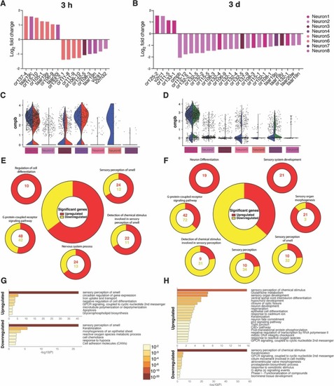
Fig. 6. Intranasal SARS-CoV-2 S RBD protein delivery causes transcriptional changes in zebrafish sustentacular and endothelial clusters. (A-B) Bar plots showing fold-change in expression of selected significantly differentially expressed genes in sustentacular and endothelial cell clusters in the zebrafish OO 3 h and 3 d post intranasal SARS-CoV-2 S RBD protein compared to vehicle controls identified by scRNA-Seq. (C-D) Summary webs of gene ontology analysis performed in ShinyGO v0.61 from the scRNA-Seq data showing the number of genes significantly up and down regulated in sustentacular and endothelial cell clusters in the OO of zebrafish 3 h and 3 days post-intranasal SARS-CoV-2 S RBD protein compared to vehicle treated controls. (E-F) Gene set enrichment analysis using Metascape of genes from sustentacular and endothelial clusters that were either significantly up or down regulated in zebrafish OO 3 h and 3 d post-intranasal SARS-CoV-2 S RBD protein compared to vehicle treated controls (adjusted P-value < 0.05). Significantly enriched terms are colored by P-value.

Acknowledgments
This image is the copyrighted work of the attributed author or publisher, and ZFIN has permission only to display this image to its users. Additional permissions should be obtained from the applicable author or publisher of the image. Full text @ Brain Behav. Immun.