
- Title
-
NAA10 p.(N101K) disrupts N-terminal acetyltransferase complex NatA and is associated with developmental delay and hemihypertrophy
- Authors
- McTiernan, N., Gill, H., Prada, C.E., Pachajoa, H., Lores, J., CAUSES study, Arnesen, T.
- Source
- Full text @ Eur. J. Hum. Genet.
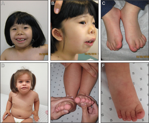
Photographs of individual 1 showing dysmorphology ( |
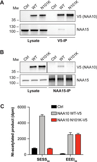
NatA complex formation and catalytic activity of immunoprecipitated NAA10 WT-V5 and NAA10 N101K-V5.
NAA10 WT-V5 and NAA10 N101K-V5 were overexpressed in HeLa cells, immunoprecipitated by V5-tag antibody (a) or NAA15 antibody (b) and analysed by Western blotting. Densitometry analysis was performed to quantify NAA10-V5 and NAA15 bands. c Nt-acetylation assay displaying catalytic activity of immunoprecipitated NAA10 WT-V5 and NAA10 N101K-V5. The measured catalytic activity toward NatA substrate SESS24 and monomeric NAA10 substrate EEEI24 was normalised to the amount of immunoprecipitated NAA15 and NAA10-V5, respectively. Reaction mixtures either with immunoprecipitated β-gal-V5 or without peptide were used as negative controls to account for background signal. The IP and activity measurements were performed in three independent setups, each with three technical replicates per assay. One representative setup is shown. |
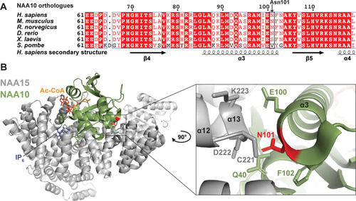
NAA10 multiple sequence alignment and NatA structural analysis.
a Multiple sequence alignment of NAA10 orthologues from human, mouse, rat, zebrafish, frog, and yeast. Secondary structure was determined from hNatA structure (PDB ID: 6C9M) [40] and amino acid conservation is indicated by red colour. b Human NatA structure (PDB ID: 6C9M) [40] with the auxiliary subunit NAA15 (grey), the catalytic subunit NAA10 (green) and Ac-CoA and IP6 shown as orange and blue sticks, respectively. The structure was superimposed on Ac-CoA from the S. pombe NAA10 structure (PDB ID: 4KVX) [13]. The variant site Asn101 is coloured red. Close-up of Asn101 shows that it is located in NAA10 α3 helix with its side chain protruding toward NAA15. |