
|
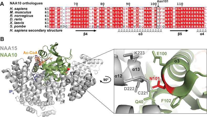
Fig. 3
a Multiple sequence alignment of NAA10 orthologues from human, mouse, rat, zebrafish, frog, and yeast. Secondary structure was determined from hNatA structure (PDB ID: 6C9M) [40] and amino acid conservation is indicated by red colour. b Human NatA structure (PDB ID: 6C9M) [40] with the auxiliary subunit NAA15 (grey), the catalytic subunit NAA10 (green) and Ac-CoA and IP6 shown as orange and blue sticks, respectively. The structure was superimposed on Ac-CoA from the S. pombe NAA10 structure (PDB ID: 4KVX) [13]. The variant site Asn101 is coloured red. Close-up of Asn101 shows that it is located in NAA10 α3 helix with its side chain protruding toward NAA15.