
- Title
-
Label-free quantitative measurement of cardiovascular dynamics in a zebrafish embryo using frequency-comb-referenced-quantitative phase imaging
- Authors
- Boonruangkan, J., Farrokhi, H., Rohith, T.M., Kwok, S., Carney, T.J., Su, P.C., Kim, Y.J.
- Source
- Full text @ J. Biomed. Opt.
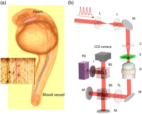
FCR-QPI. (a) Zebrafish embryo imaged under a WL microscope ( |
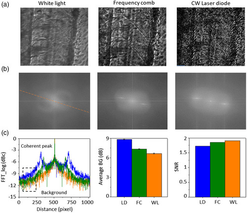
Evaluation of imaging performance of different light sources in QPI. (a) QPI images of zebrafish trunk illuminated by WL, FC, and continuous-wave LD. (b) FFT spectrums analyzed from the QPI images in (a). (c) Line extraction from the FFT spectrums of three light sources. (d) Average background (BG) noise and SNR, calculated in the Fourier domain. |

Phase reconstruction of an RBC from QPI interferogram. (a) Procedures for phase reconstruction of a single RBC. (b) Time-lapse phase maps of RBC flow in the blood vessel. |
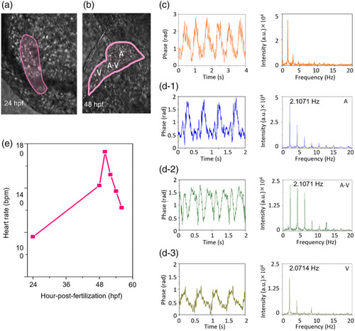
FCR-QPI for measuring the dynamic motions of zebrafish’s heart. Phase images of zebrafish’s heart at (a) 24 hpf and (b) 48 hpf. (c) Periodic signals of the heartbeat at 24 hpf analyzed in time and frequency domains. Periodic signals of the heartbeat at 48 hpf measured at (d-1) atrium (A), (d-2) A-V canal, and (d-3) ventricle (V). (e) Heart rate measured at various hpf. |

Blood flow measurement. (a) Schematic drawing of RBCs flowing in the blood vessels at 24 hpf. (b) Signal of RBCs flowing in the DA. (c) Mean RBC velocity in DA and PCV. (d) Maximum RBC velocity in DA and PCV. (e) Schematic drawing of RBCs flowing in the blood vessels at 48 hpf. Signal of pulsatile RBCs flowing in the (f) DA and (g) PCV. (h) Conceptual drawing of the number of RBCs and pulse ratio. (i) Number of RBCs and (j) pulse ratio in one blood pulse measured in DA, CA, PCV, and CV. DA, dorsal aorta; CA, caudal artery; PCV, post-cardinal vein; CV, caudal vein. |