FIGURE SUMMARY
Title

Exercise, programmed cell death and exhaustion of cardiomyocyte proliferation in aging zebrafish

Authors
Murphy, L.B., Santos-Ledo, A., Dhanaseelan, T., Eley, L., Burns, D., Henderson, D.J., Chaudhry, B.
Source
Full text @ Dis. Model. Mech.

Zebrafish do not exhibit indeterminate somatic or ventricular growth. (A) External appearances of male and female zebrafish between 6 months and 36 months of age. Female fish have larger abdomen than male fish (asterisk). Spinal deformities are more common and increasingly severe after 24 months (arrows). (B) Lengths of male (blue points) and female (red points) zebrafish with increasing age. Sample size (n) is 11, 11, 10, 11, 6 for age groups 6, 12, 18, 24 and 36 months, respectively. There were no differences between the lengths of male and female fish at any stage. After a rapid increase in length in the first 6 months, there was a smaller increase in length in the second 6 months of life, but no change thereafter. (C) Midline histological sections through zebrafish hearts stained with Masson's trichrome. Myocardium is indicated in brown and collagen in blue. The 6-month-old heart had minimal collagen staining in the bulbus arteriosus (ba). Epicardial adipose tissue is not seen. At 12 months, the heart was larger and the bulbus stained strongly for collagen fibres, and there is some epicardial adipose tissue (arrowheads). In the 36-month-old heart, there were extensive epicardial adipose deposits seen on the surface of the heart (arrowheads). There were also extensive collagen deposits in the bulbus arteriosus (ba), bulbo-ventricular valve (bvv) and atrioventricular valve (avv). (D) Ventricular length measured from the histological sections in C, from apex to base of chamber, indicated by the dashed bar in C (6 months). All individual data points are shown (male, blue points; female, red points). Sample size (n) was 11, 11, 10, 11 and 6 for age groups 6, 12, 18, 24 and 36 months, respectively. Ventricular length increased between 6 and 12 months but not thereafter, and there was no difference between hearts from male and female fish. (E) Mean thickness of the ventricular myocardium compact layer increased until 18 months of age and did not change thereafter. Sample size (n) was 5, 5, 7, 7 and 5 for age groups 6, 12, 18, 24 and 36 months, respectively. (F) Ratio of ventricular length (D) to body length (B). Sample size (n) is 10, 11, 9, 11 and 6 for age groups 6, 12, 18, 24 and 36 months, respectively. There was no difference in the ratio throughout adult life. All individual data points are shown. Data are mean±s.e.m. *P<0.05; **P< 0.02; ns, not significant (two-way ANOVA was used in B,D; one-way ANOVA in E,F). a, atrium; avv, atrioventricular valve; ba, bulbus arteriosus; bvv, bulbo-ventricular valve; v, ventricle.

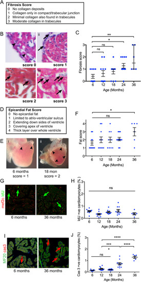
(A) Description of semiquantitative fibrosis score. (B) Appearances of each fibrosis score category. Arrowheads (i) indicate the absence of collagen at the compact-trabecular junction. Arrows indicate deposits of fibrosis at the compact-trabecular junction (ii) and in the trabecular myocardium (iii and iv). (C) Fibrosis score with age. Sample size (n) was 10, 11, 10, 11 and 6 for age groups 6, 12, 18, 24 and 36 months, respectively. All individual data points are shown with median and quartiles. The median fibrosis score was elevated at 24 and 36 months (Kruskal–Wallis test, non-parametric ANOVA). (D) Description of semiquantitative epicardial fat score. (E) Epicardial fat limited to the atrioventricular sulcus (i) and extending over sides (ii). (F) Increased epicardial fat score at 36 months (one-way ANOVA). Sample size (n) was 10, 11, 10, 11 and 6 for age groups 6, 12, 18, 24 and 36 months, respectively. (G) Examples of immunofluorescence labelling of IdU incorporation (arrows) at 6 and 36 months. (H) Percentage of cardiomyocytes incorporating IdU remains constant with age. Sample size (n) was 10, 10, 10, 10 and 6 for age groups 6, 12, 18, 24 and 36 months, respectively. (I) Nuclei labelling with activated caspase 3 antibodies (arrows). (J) Increased frequency of caspase+ cardiomyocyte nuclei from 24 months of age (one-way ANOVA). Sample size (n) was 10, 11, 10, 11 and 6 for age groups 6, 12, 18, 24 and 36 months, respectively. All individual data points are shown. Data are mean±s.e.m. *P<0.05; **P<0.02; ***P<0.001; ****P<0.0001; ns, not significant.

Physiological effects of aging in zebrafish. (A) Experimental setup to assess voluntary swimming. Individual fish were placed in a 3-litre tank, with white paper below and digital video captured from above. The position of the head was tracked manually using Fiji/ImageJ. (B) Voluntary swimming speeds in body lengths per second for 6-month and 18-month-old zebrafish. Sample size (n) was ten for both 6- and 18-month age groups. (C,D) Quantitative RT-PCR analysis of nppa and nppb gene expression in whole hearts. Sample size (n) was ten for both 6- and 18-month age groups. All individual data points are shown, mean±s.e.m. Normal distribution of data (D'Agostino–Pearson), no difference in variance (F-test). *P<0.05; **P<0.02; ns, not significant (unpaired two-tailed Student's t-test).

Critical swimming speed diminishes with aging. (A) Critical swimming speed protocol. Individual fish were placed in a modified flume containing a narrow cross-section to increase water speed. During the first 30 min, the water speed is increased to 14 cm sec−1 to allow acclimatisation and allow transition from dart and glide to sustained swimming patterns. When the fish were unable to maintain swimming speed, the water speed of the last completed stage, with allowance for proportion of current stage, was used to calculate Ucrit. A flow meter (L min−1) was used to follow increases in water speed (cm sec−1). (B) Progressive fall in Ucrit from 6 to 21 months (one-way ANOVA). All individual data points are shown, Data are mean±s.e.m. Sample size (n) was 7, 13, 26, 6 and 3 for age groups 6, 18, 21, 24 and 36 months, respectively. ***P<0.001; ****P<0.0001; ns, not significant.

Cardiomyocyte turnover in response to 3-day high-intensity swimming stress test. (A) Experimental protocol. Zebrafish were first exposed to water containing IdU for 24 h. They were then placed in the Ucrit flume, modified to house groups of fish at moderate water flow by removing the narrow channel insert. The exercise group were placed in running water at 3 BL sec−1 for 72 h. The control groups were placed in an identical flume with no water flow (rest conditions). After 72 h in the flumes, all fish received 12 h of rest in holding tanks and were exposed to CldU for 24 h, and then culled. (B) Percentage of cardiomyocytes labelling with IdU or CldU in 7-month-old fish under high-intensity swimming (n=10) or rest conditions (n=10). (C) Percentage of cardiomyocytes labelled with IdU or CldU in 21-month-old fish under high-intensity swimming (n=7) or rest conditions (n=5). (D) Percentage of cardiomyocytes dual labelled with IdU and CldU in 7-month-old and 21-month-old zebrafish under rest and high-intensity swimming conditions. Sample numbers as in B and C. (E) Venn diagrams summarising CldU and IdU results. Diameter of circles indicates relative magnitude of thymidine analogue incorporation. Percentages indicate proportions of cycling cardiomyocytes within each group. (F) Percentage of cardiomyocytes immunolabelling with activated caspase 3 antibodies in 7-month-old zebrafish under rest (n=9) or exercise (n=8), and 21-month-old zebrafish under rest (n=10) or exercise (n=8) conditions. Data are mean±s.e.m. **P<0.02; ****P<0.0001; ns, not significant (one-way ANOVA).

Cardiomyocyte turnover in response to 3-day high-intensity swimming stress test after being raised in running water. (A) Experimental protocol. Data from fish raised under normal water conditions (normal) are reproduced from Fig. 5 for ease of comparison. Long-term running water conditions were produced by placing an 8-litre min−1 impeller pump/filter unit inside a holding tank containing siblings of fish described in Fig. 5 when 10 months of age. Both normal and running water conditions zebrafish were given the 3-day high-intensity swimming stress test (see Fig. 5), and critical swimming speed was then determined (see Fig. 4). (B) Critical swimming speed of zebrafish raised in normal (n=15) and in running water conditions (n=12) at 21 months. No difference in variance. (C) IdU and CldU incorporation into cardiomyocytes for zebrafish raised in normal conditions (n=5) and in running water (n=6) with the 3-day high-intensity swimming protocol under rest conditions. (D) IdU and CldU incorporation into cardiomyocytes for zebrafish raised in normal (n=7) and running water (n=5) with the 3-day high-intensity swimming protocol under exercise conditions. (E) Percentage of cardiomyocytes dual labelled with both IdU and CldU under rest or exercise conditions for zebrafish raised in normal or running water conditions. Subject numbers as in C and D. (F) Venn diagrams summarising CldU and IdU results. Diameter of circles indicates relative magnitude of thymidine analogue incorporation. Percentages indicate proportions of cycling cardiomyocytes within each group. (G) Percentage of cardiomyocyte immunolabelling with activated caspase 3 antibodies in 21-month-old zebrafish raised in normal conditions under rest (n=5) or exercise (n=7), and for zebrafish raised in running water conditions under rest (n=6) or exercise conditions (n=5). Data are mean±s.e.m. Statistical significance was determined using an unpaired two-tailed Student's t-test (B) and one-way ANOVA (C-E,G). *P<0.05; **P<0.02; ns, not significant.

Increased cardiac fibrosis after prolonged exercise. (A) Body length in 21-month-old fish raised in either normal (n=13) or running water (n=12). (B) Ventricle length in 21-month-old fish raised in either normal (n=12) or running water (n=11). (C) Increased myocardial fibrosis in zebrafish maintained in running water conditions. (D) Myocardial fibrosis scores in zebrafish maintained in running water conditions (n=12) or running water (n=11). Data are mean±s.e.m. *P<0.05; ns, not significant (unpaired two-tailed Student's t-test).

Acknowledgments
This image is the copyrighted work of the attributed author or publisher, and ZFIN has permission only to display this image to its users. Additional permissions should be obtained from the applicable author or publisher of the image. Full text @ Dis. Model. Mech.