
- Title
-
Zebrafish stm is involved in the development of otoliths and of the fertilization envelope
- Authors
- Pachoensuk, T., Fukuyo, T., Rezanujjaman, Md., Wanlada, K., Yamamoto, C., Maeno, A., Mostafizur Rahaman, Md., Hasan Ali, Md., Tokumoto, T.
- Source
- Full text @ RAF
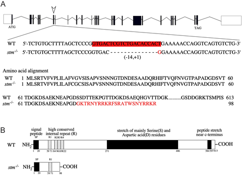
DNA sequence and predicted protein structure of an established stm gene-edited strain. (A) Genome structure and DNA sequence of the target site for the genome editing of stm. DNA sequences around the target site (in red) for CRISPR/Cas9 digestion in WT and stm−/− mutants are indicated. A 14-nucleotide deletion and 1-nucleotide insertion were induced in the target site in the selected mutant. (B) The predicted protein structures of the WT and the stm−/− mutant are indicated. It is expected that a peptide of 98 amino acids in length with an N-terminal 76-amino acid sequence is the same as WT Stm and is produced in the stm−/− mutant. Thus only first high conserved internal repeat (R1) is present in Stm protein produced in the stm−/− mutant. |
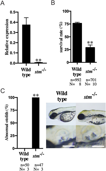
Expression of stm, survival rate and abnormalities of otoliths in stm−/− mutant. (A) Relative expression of stm in embryos of WT and stm−/− mutant were compared. mRNA abundance was measured in triplicate for each sample from three different paring, and all data were normalized by the number of elongation factor 1α (EF1α) transcripts in each sample. (B) Survival rates of embryos in the WT and stm−/− mutant (F6 generation) at 5 dpf. n = total number of embryos; N = number of biological replicates (embryos from different pair of fishes). (C) Percentages of abnormal otolith containing embryos in the WT and stm−/− mutant. n = total number of embryos; N = number of biological replicates (embryos from different pair of fishes). Representative otoliths of 5 dpf embryos in WT and stm−/− mutant are indicated. Scale bars are 100 µm. Asterisks represent significant difference between the samples (**P ≤ 0.001). PHENOTYPE:
|
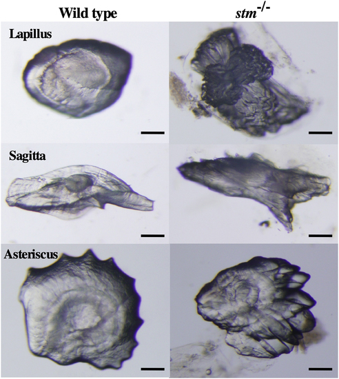
Stereomicroscopic observation of otoliths. Photographs of three excised otoliths (Lapillus, Sagitta and Asteriscus) from adult WT zebrafish and the stm−/− mutant are indicated. Scale bars are 100 µm. PHENOTYPE:
|
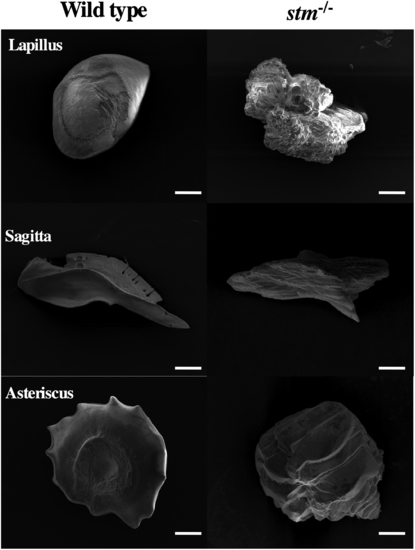
Observation of otoliths by SEM. Photographs from scanning electron microscopy observations of three excised otoliths (Lapillus, Sagitta and Asteriscus) from adult WT zebrafish and the stm−/− mutant are indicated. Scale bars are 100 µm. PHENOTYPE:
|
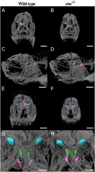
Observation of otoliths by micro CT. Micro CT scan images from anterior (A and B), left lateral (C and D), posterior (E and F) and dorsal (G and H) sides of adult WT zebrafish and the stm −/− mutant are indicated. Scale bars in A–F are 1 mm. Scale bars in G and H are 500 µm. Three otoliths are indicated in different colours: Lapillus; blue, Sagitta; green, Asteriscus; magenta. Scale bars are 400 µm. PHENOTYPE:
|
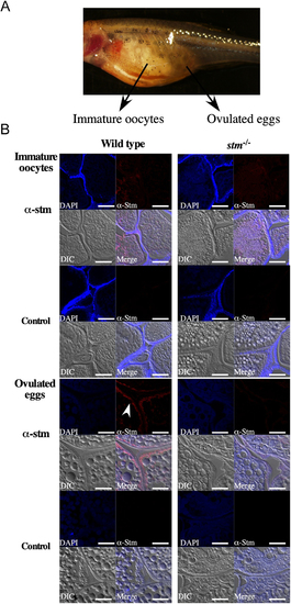
Immunohistochemical observation of Stm. (A) A photograph of stm−/− mutant females possessing ovulated eggs. Frozen sections of immature oocytes and ovulated eggs were prepared from the fish. (B) Immunohistochemical staining results for Stm in WT and in stm−/− mutant immature oocytes (upper panel) and ovulated eggs (lower panel). Frozen sections of the whole bodies of WT and stm−/− mutant zebrafish females were stained with anti-Stm antibodies (α-stm) and DAPI. Differential contrast (DIC) images and merged images of anti-Stm, DAPI and DIC are also indicated (Merge). Control staining (Control) without anti-Stm antibodies of each sample is indicated below. The white arrow indicates signals in anti-Stm staining. The scale bars indicate 50 µm. PHENOTYPE:
|
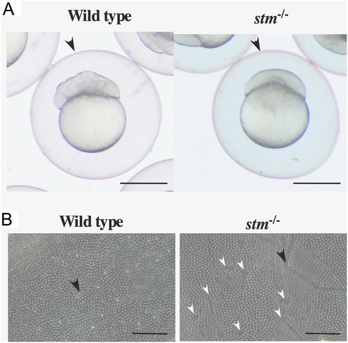
Observation of the FE. (A) Photographs of FE-covered embryos of WT and stm−/− mutants. FEs are indicated by arrowheads. Scale bars are 500 µm. (B) Photographs from microscope observation of FE from WT and stm−/− mutant embryos are indicated. A crystal-like structure is indicated by the black arrow. Cracks in the FE of stm−/− mutants are indicated by white arrowheads. Scale bars are 20 µm. PHENOTYPE:
|
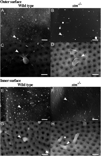
Observation of the FE by SEM. Photographs from scanning electron microscopy observations of the outer surface of the FE (A and B in 1500×; C and D in 8000×) and the inner surface of the FE (E and F in 1500×; G and H in 8000×) from adult WT zebrafish and the stm−/− mutant are indicated. A fibre-supported knob-like structure is indicated by an arrow. Scale bars in A, B, E, F are 2 µm. Scale bars in C, D, G, H are 10 µm. |
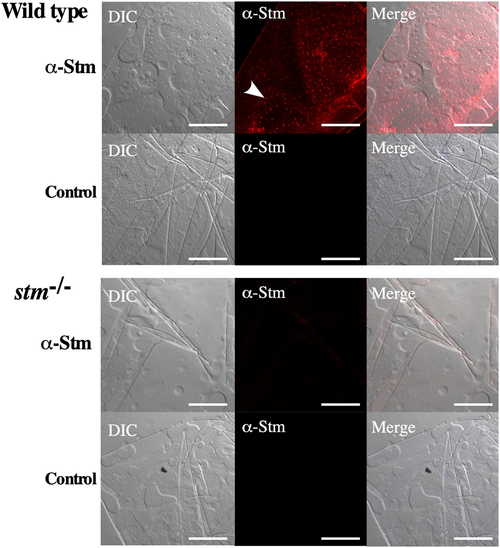
Immunohistochemical observation of Stm on FE. Immunohistochemical staining results for Stm of FE from WT and in stm−/− mutant. FEs of WT and stm−/− mutant zebrafish embryos were stained with anti-Stm antibodies (α-Stm). Differential contrast (DIC) images and merged images of anti-Stm and DIC are also indicated (Merge). Control staining (Control) without anti-Stm antibodies of each sample is indicated below. The white arrow indicates signals in anti-Stm staining. The scale bars indicate 50 µm. |

Unillustrated author statements PHENOTYPE:
|