FIGURE SUMMARY
Title

Association Analysis of Variants of DSCAM and BACE2 With Hirschsprung Disease Susceptibility in Han Chinese and Functional Evaluation in Zebrafish

Authors
Lu, Y.J., Yu, W.W., Cui, M.M., Yu, X.X., Song, H.L., Bai, M.R., Wu, W.J., Gu, B.L., Wang, J., Cai, W., Chu, X.
Source
Full text @ Front Cell Dev Biol

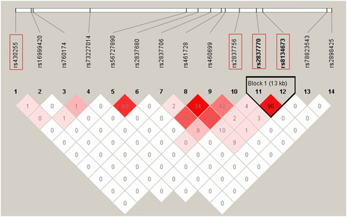
Linkage disequilibrium (LD) pattern of 12 DSCAM single-nucleotide polymorphisms (SNPs) with P < 0.05 in association analysis and two previous reported SNPs. The plot was constructed using the program Haploview, and r2 values (×100) between SNPs are shown in the diamonds. r2 values were calculated using data of 420 HSCR patients and 1,663 controls from the current study. r2 values of 1 represent complete LD, r2 values greater than 0.8 represent strong evidence of LD, r2 values of 0.2–0.8 represent moderate LD, and r2 values less than 0.2 represent low LD. Haplotype blocks were determined using the confidence interval method. Two SNPs (rs2837770 and rs8134673) reported associations with isolated HSCR in previous study, and the two most associated SNPs (rs430255 and rs2837756) are highlighted in the red rectangles.

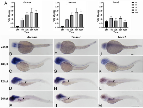
Spatiotemporal expression of zebrafish of dscama, dscamb, and bace2 during early embryogenesis. (A) qRT-PCR analysis of relative expression levels of dscama, dscamb, and bace2 in zebrafish embryos from 24 to 120 hpf. (B–M) Whole-mount in situ hybridization in wild-type embryos for dscama(B–E), dscamb(F–I), and bace2(J–M). Strong expression of dscama(B–E) and dscamb(F–I) was observed in the brain and central nervous system after 24 hpf, and a weakly positive expression signal was observed in the intestinal bulb at 72 and 96 hpf (black arrowheads). (J–K)bace2 expression in the neural crest cells and pigment epithelial cells of the retina at 24 and 48 hpf. A relatively strong expression signal of bace2 was observed in the intestinal primordium after 72 hpf indicated by black arrowheads (L,M). Scale bar for 24 and 48-hpf embryos is 100 μm, and that for 72 and 96-hpf embryos is 500 μm.

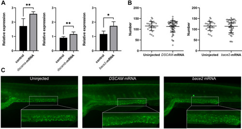
Phenotypes of zebrafish embryos with overexpression of
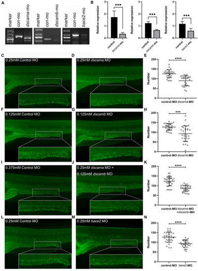
 DSCAM and bace2. Zebrafish embryos were injected with 100 pg of DSCAM mRNA per embryo and 100 pg of bace2 mRNA per embryo, respectively. (A) The expression levels of dscama, dscamb, and bace2 of mRNA injected embryos were analyzed by qRT-PCR at 48 hpf (P < 0.05, ∗∗P < 0.01). (B) No significant difference was found in the neuron numbers of the last six somite lengths of gut between control and mRNA injected embryos at 5 dpf. The HuC/D antibody was used to stain the enteric nervous system (ENS) neurons, and the neuron numbers in the last six somite lengths of gut were counted. Embryos injected with DSCAM mRNA, n = 67. Embryos injected with bace2 RNA, n = 53. Control embryos, n = 28. (C) The overexpression of DSCAM and bace2 caused no obvious defect of enteric neurons in the hindgut.

Phenotypes of dscama, dscamb, and bace2 knockdown zebrafish. Embryos at 5 dpf were stained with HuC/D antibody to observe the intestinal neurons. The neuron numbers in the last six somite lengths of gut were counted. (A) RT-PCR confirmation of splice-blocking morpholino oligonucleotide (SBMO) knockdown in embryos at 48 hpf. In dscama morphants, two small fragments were observed. In dscamb morphants, a shorter fragment was produced, and the amount of PCR products decreased. The length of PCR products from mRNA of bace2 morphants was same as control MO-injected embryos, but the amount of PCR product decreased obviously. (B) qRT-PCR analysis of dscama, dscamb, and bace2 mRNA expression after morpholino injection. The target gene expression levels of SBMO-injected embryos were remarkably reduced compared with the controls. (C–E) Compared with the 0.25 mM control MO injection, 0.25 mM dscama MO injection resulted in a decrease in the density of enteric neurons. (F–H) Embryos injected with 0.125 mM of control MO and 0.125 mM of dscamb MO. (I–K) Embryos injected with 0.375 mM of control MO and 0.25 mM of dscama MO + 0.125 mM of dscamb MO. (L–N) Embryos injected with 0.25 mM of control MO and 0.25 mM of bace2 MO. ∗∗∗P < 0.001, *⁣*⁣**P < 0.0001.

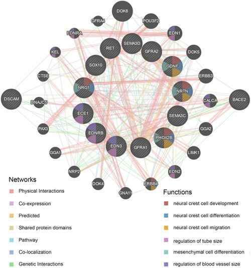
Protein–protein interaction (PPI) network of DSCAM and BACE2. The plot was constructed on GeneMANIA website. The correlations of DSCAM and BACE2 with critical signaling pathway genes were illustrated. The 13 central nodes represent the critical susceptibility genes of HSCR. The surrounded 20 nodes represent genes highly relevant to the central nodes in terms of physical interaction, co-expression, prediction, pathway, co-localization, genetic interactions, and shared protein domains. The color of connecting lines between nodes represents the type of protein–protein interaction (PPI). The color of the node represents the possible functions of the genes.

Acknowledgments
This image is the copyrighted work of the attributed author or publisher, and ZFIN has permission only to display this image to its users. Additional permissions should be obtained from the applicable author or publisher of the image. Full text @ Front Cell Dev Biol