FIGURE SUMMARY
Title

Fisetin inhibits lipopolysaccharide-induced inflammatory response by activating β-catenin, leading to a decrease in endotoxic shock

Authors
Molagoda, I.M.N., Jayasingha, J.A.C.C., Choi, Y.H., Jayasooriya, R.G.P.T., Kang, C.H., Kim, G.Y.
Source
Full text @ Sci. Rep.

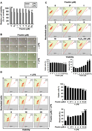
High concentrations of fisetin decreases the cell viability of RAW 264.7 macrophages. RAW 264.7 macrophages (1 × 105 cells/mL) were treated with fisetin (0–20 µM) for 24 h in the presence or absence of 500 ng/mL LPS. (A) An MTT assay was performed. (B) Microscopic images was taken using phase contrast microcopy (× 10). Scale bares 40 μm. (C,D) Total viable cells and dead cells in the absence (C) and presence (D) of LPS were determined by flow cytometry. Each value indicates the mean ± SEM from three independent experiments. Significant differences among the groups were determined using an unpaired one-way ANOVA with Bonferroni correction. ###p < 0.001 and #p < 0.05 vs. untreated cells; ***p < 0.001 vs. LPS-treated cells. 0v, vehicle control (0.1% DMSO).

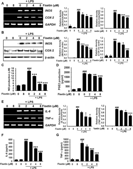
Fisetin decreases LPS-induced inflammatory mediator and cytokine levels in RAW 264.7 macrophages. RAW 264.7 macrophages (1 × 105 cells/mL) were treated with fisetin (0–8 µM) 2 h before treatment with 500 ng/mL LPS. (A) Total mRNA was isolated at 6 h after 500 ng/mL LPS treatment, and RT-PCR was performed. GAPDH was used as an internal control. (B) Total proteins were isolated at 24 h and western blotting was performed. β-Actin was used as an internal control. (C) The amount of NO production in the culture medium was determined using the Griess Reagent Assay. (D) The amount of PGE2 was determined at 24 h using an ELISA according to the manufacturer’s instructions. (E) Total mRNA was isolated at 6 h and subjected to RT-PCR for IL-6 and TNF-α. The amount of (F) IL-6 and (G) TNF-α was measured at 24 h by an ELISA. Each value indicates the mean ± SEM from three independent experiments. Significant differences among the groups were determined using the Student’s t-test (###p < 0.001 vs. untreated cells) and one-way ANOVA with Bonferroni correction (***p < 0.001, **p < 0.05, and *p < 0.01 vs. LPS-treated cells).

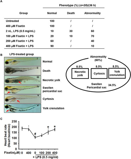
Fisetin attenuates LPS-induced mortality, abnormalities, and lowered heart rate in zebrafish larvae. Zebrafish larvae at 3 dpf (n = 20) were microinjected with 2 nL of 0.5 mg/mL LPS and immediately immersed in the indicated concentrations of fisetin. (A) Phenotype-based evaluations were performed at 36 h post injection (hpi). (B) LPS-microinjection increases abnormalities in zebrafish larvae at 36 hpi; this was observed using stereomicroscopy; (i) normal, (ii) death, (iii) necrotic yolk, (iv) swollen pericardial sac, (v) cyrtosis, and (vi) yolk crenulation. (C) LPS microinjection induces 60% abnormalities of zebrafish larvae and all larvae exhibits swollen pericardial sac (34.5%) either alone or together with necrotic yolk (8.5%), cyrtosis (8.5%), or yolk crenulation (8.5%). (D) Heart rates were measured to assess toxicity. Each value indicates the mean ± standard error median (SEM), and is representative of the results obtained from 20 fish for each group. Significant differences among the groups were determined using Student’s t-test (#p < 0.01 vs. untreated zebrafish larvae) and one-way ANOVA with Bonferroni correction (*p < 0.01 vs. LPS-treated zebrafish larvae).

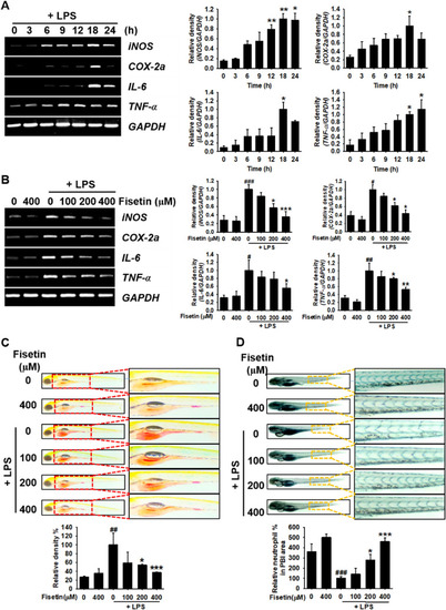
Fisetin inhibits LPS-induced inflammatory response in zebrafish larvae. Zebrafish larvae at 1 day post fertilization (dpf) were cultured in 0.003% PTU containing E3 embryo media. Briefly, 2 nL of 0.5 mg/mL LPS was microinjected into the yolk at 3 dpf. Zebrafish larvae were immediately immersed in E3 embryo media containing different concentrations of fisetin. (A) In LPS-microinjected conditions, 10 zebrafish were euthanized at the indicated time points and subjected to RT-PCR for evaluating the expression of iNOS, COX-2a, IL-6, and TNF-α. (B) At 18 h post injection (hpi), 20 zebrafish larvae from each treatment were euthanized and the expression of iNOS, COX-2a, IL-6, and TNF-α was measured by RT-PCR. (C) Neutral red staining of macrophages and (D) sudan black staining of the neutrophils were performed at 24 hpi. Each value indicates the mean ± standard error median (SEM) and is representative of the results obtained from three independent experiments. Significant differences among the groups were determined using the Student’s t-test (###p < 0.001, ##p < 0.01, and #p < 0.05 vs. untreated zebrafish larvae) and one-way ANOVA with Bonferroni correction (***p < 0.001, **p < 0.01, and *p < 0.05 vs. LPS-treated zebrafish larvae).

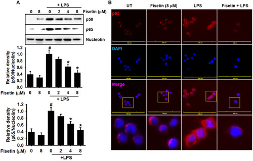
Fisetin inhibits LPS-induced nuclear translocation of NF-κB in RAW 264.7 macrophages. RAW 26.4.7 cells (1 × 105 cells/mL) were pretreated with fisetin 2 h before LPS treatment (500 ng/mL; 0.5 h). (A) Nuclear proteins were isolated and equal amounts of proteins were resolved on SDS–polyacrylamide gels, transferred to PVDF membranes, and probed with antibodies against p50 and p65. Nucleolin was used as an internal control. (B) The nuclear translocation of p65 was detected by fluorescence microscopy. Each value indicates the mean ± standard error median (SEM), and is representative of the results obtained from three independent experiments. Significant differences among the groups were determined using Student’s t-test (#p < 0.05 vs. the untreated group) and one-way ANOVA with Bonferroni correction (*p < 0.05 vs. the LPS-treated cells). UT untreatment.

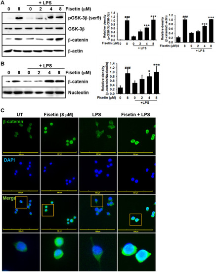
Fisetin promotes the expression of β-catenin and its nuclear translocation in LPS-stimulated RAW 264.7 macrophages. RAW 264.7 macrophages (1 × 105 cells/mL) were pretreated with fisetin 2 h prior to LPS treatment (500 ng/mL; 0.5 h). (A) Total proteins were isolated and equal amounts of proteins were resolved on SDS–polyacrylamide gels, transferred to PVDF membranes, and probed with antibodies against total GSK-3β, phosphor-GSK-3β (Ser9), and β-catenin. β-Actin was used as an internal control. (B) In a parallel experiment, nuclear proteins were isolated and western blotting was performed to verify the nuclear translocation of β-catenin. Nucleolin was used as an internal control. (C) Nuclear translocation of β-catenin was detected by immunofluorescence. Each value indicates the mean ± standard error median (SEM), and is representative of the results obtained from three independent experiments. Significant differences among the groups were determined using Student’s t-test (###p < 0.001 vs. untreated cells) and one-way ANOVA with Bonferroni correction (***p < 0.001 and *p < 0.01 vs. LPS-treated cells). UT untreatment.

Fisetin inhibits LPS- or FH535-mediated IL-6 and TNF-α release by stimulating β-catenin-mediated NF-κB inactivation. RAW 264.7 macrophages (1 × 105 cells/mL) were incubated with 10 µM FH535 for 2 h prior to treatment with 8 μM fisetin and 500 ng/mL LPS. (A) Nuclear proteins were extracted and western blotting was performed with specific antibodies against p65, p50, and β-catenin. Nucleolin was used as an internal control. (B,C) In a parallel experiment, the culture supernatants were collected and extracellular levels of (B) IL-6 and (C) TNF-α were measured by ELISA. Each value indicates the mean ± standard error median (SEM), and is representative of the results obtained from three independent experiments. Significant differences among the groups were determined using one-way ANOVA with Bonferroni correction (***p < 0.001, **p < 0.005, and *p < 0.01).

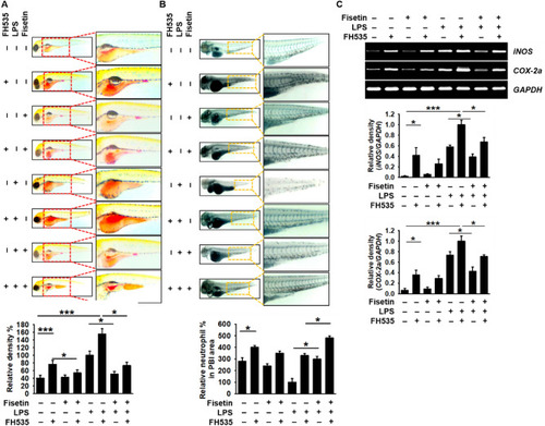
Fisetin inhibits LPS-induced inflammation and endotoxic shock by stimulating the β-catenin signaling pathway in zebrafish larvae. Zebrafish larvae at 3 dpf were pretreated with 10 µM FH535 for 2 h prior to LPS (2 nL of 0.5 mg/mL) microinjection. Then, the larvae were grown in E3 media containing 400 μM fisetin. (A) Neutral red-stained macrophages are shown at 18 hpi. (B) Sudan black-stained neutrophils were observed at 18 hpi. (C) At 18 hpi, 10 zebrafish from each condition were collected and the expression of iNOS and COX-2a was detected by RT-PCR. GAPDH was used as an internal control. Each value indicates the mean ± standard error median (SEM) and is representative of the results obtained from three independent experiments. Significant differences among the groups were determined using one-way ANOVA with Bonferroni correction (***p < 0.001, **p < 0.005, and *p < 0.01).

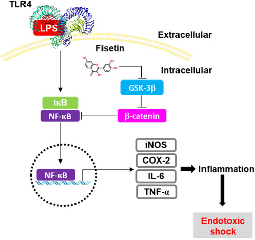
Fisetin inhibits GSK-3β-mediated NF-κB activation in the presence of β-catenin, leading to the inhibition of inflammation-induced septic shock. Once macrophages are exposed to high concentrations of the bacterial endotoxin, LPS, they initiate an inflammatory response and endotoxic shock by upregulating the expression of NF-κB-induced inflammatory genes, such as iNOS, COX-2, IL-12, IL-6, and TNF-α. Fisetin binds to the noncompetitive ATP-binding sites of GSK-3β and phosphorylates GSK-3β at Ser9, resulting in the inactivation GSK-3β and release of β-catenin from the destruction complex. The released β-catenin inhibits the transcriptional activity of NF-κB, thereby alleviating LPS-induced inflammation and endotoxic shock.

Acknowledgments
This image is the copyrighted work of the attributed author or publisher, and ZFIN has permission only to display this image to its users. Additional permissions should be obtained from the applicable author or publisher of the image. Full text @ Sci. Rep.