FIGURE SUMMARY
Title

A hypomorphic variant in EYS detected by genome-wide association study contributes toward retinitis pigmentosa

Authors
Nishiguchi, K.M., Miya, F., Mori, Y., Fujita, K., Akiyama, M., Kamatani, T., Koyanagi, Y., Sato, K., Takigawa, T., Ueno, S., Tsugita, M., Kunikata, H., Cisarova, K., Nishino, J., Murakami, A., Abe, T., Momozawa, Y., Terasaki, H., Wada, Y., Sonoda, K.H., Rivolta, C., Tsunoda, T., Tsujikawa, M., Ikeda, Y., Nakazawa, T.
Source
Full text @ Commun Biol

Genome-wide association study (GWAS) of ARRP patients and detection of three independent signals in <italic>EYS</italic>.

a Results of a meta-GWAS displayed as a Manhattan plot. Genome-wide significance (P = 5.0 × 10−8) and possible significance (P = 1.0  ×  10−5) are marked with red and blue lines, respectively. A single peak at the EYS locus surpassed genome-wide significance. b Results of a conditional analysis presented as a regional plot. Three independent peaks at P < 5.0 × 10−8 were delineated after conditioning (Peaks 1–3). c Linkage disequilibrium plot using all non-synonymous variants (identified in >5% of cases) and lead variants for Peaks 1–3 identified in GWAS in presumed ARRP patients. The linkage disequilibrium plot was generated using Haploview (ver. 4.1). The default color setting of the software was used for block color setting (D′/LOD). The numbers on the blocks indicate r2 × 100; numbers are shown on the blocks only for pairs with r2 > 0.3. Peaks 1, 2, and 3 were in linkage disequilibrium with G843E, S2556C, and S1635Kfs, respectively. The lead variants for Peaks 1–3 are shown in red. Reported pathogenic founder mutations11 are shown in green, while non-synonymous variants linked to the lead variants are shown in blue. Note, S1653Kfs, a reported founder mutation linked to a GWAS lead variant, was shown in green.

Degradation of the <italic>EYS</italic> G843E mutant mRNA in patient-derived lymphoblastoid cell lines (LCLs).

a A schematic map of the RT-PCR primer designed in relation to the exon–intron structure and mutations (G843E and S1653Kfs) in EYS and published transcript variants (Tv)25. The locations of G843E (Exon 16) and S1653Kfs (Exon 26) are indicated by the arrows. Exon numbers are based on Tv1. Note, Tv5 was identified only in fibroblasts25. b RT-PCR analysis. The regions for exons 5–6, exons 14–18, and exons 40–43 of EYS were amplified on cDNA generated from patient-derived lymphoblast cell lines with wild-type EYS (normal), homozygous S1653Kfs, and homozygous G843E. The Y79 retinoblastoma cell line was used as a positive control. Note C-terminal exons of the long isoform Tv1 were detected in LCLs with homozygous G843E but not in that with homozygous S1653Kfs. Sanger sequencing of the RT-PCR amplicon confirmed the expression of the G843E mutation using a primer pair targeting exons 14–18. Meanwhile, mRNA for exons 4–5 and 14–18 were detectable, possibly reflecting the differential expression of distinct EYS isoforms25. c Chromatogram for RT-PCR amplicon (exons 14–18). Note, G843E variant is present in the patient’s mRNA. d RT-PCR analysis after mutation replacement genome-editing treatment (GE) or inhibition of nonsense-mediated mRNA decay (NMD) in LCL from an S1653Kfs homozygote, after which expression of exons 40–43 was detected.

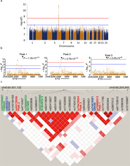
a Domain structure of EYS in relation to G843E. b Conservation of G843 across diverse species ranging from zebrafish to humans. The multiple sequence alignment was generated using ClustalW. Accession numbers of the protein sequences used for sequence comparison are as follows: human, NM_001142800.1; macaque, XM_011737495.1; pig, XM_021084496; chicken, XM_015284845.1; zebrafish, XM_009307513.

Functional assessment of <italic>EYS</italic> G843E variant following morpholino-mediated knockdown of <italic>eys</italic> in zebrafish.

a Immunostaining of Eys (green) in zebrafish retina at 1-year post-fertilization (ypf). b High-magnification image of photoreceptors. Eys (arrowhead, green) localized at the basal side of connecting cilium (acetylated α tubulin, red) of the photoreceptors. cf Expression of Eys during development at 3 days post- post-fertilization (pdf), 4, 5, and 6 dpf. g RT-PCR of eys at 4 dpf (45 cycles) following injection of three different MOs. h Quantitative RT-PCR analyses of the morphants (biologically independent samples). SP1MO (N = 5), SP2MO (N = 3), and SP3MO (N = 3). Eys expression was reduced by at least 50% at 4 dpf. Vertical bar: mean ± standard deviation. il Basal intracellular deposition of rhodopsin (rhodopsin mislocalization) observed following injection of three different MO at 6 dpf. Note, injections of three different MOs resulted in the same phenotype. m, n Rhodopsin localization in the photoreceptors at 7 dpf. m Rhodopsin is correctly localized at the photoreceptor outer segments in the control. neys knockdown by MO-induced rhodopsin mislocalization toward the basal and the lateral membrane of the photoreceptors (N = 6 biologically independent fishes). o, p Greater improvement of the rhodopsin mislocalization was achieved in the eyes supplemented with wild-type human EYS mRNA (p) over those injected with mutant human EYS mRNA with G843E (o) after SPMO2-mediated knockdown of eys, consistent with decreased EYS function by the mutation. q A quantitative analysis of o (N = 9 biologically independent fishes) and p (N = 9 biologically independent fishes). Numbers of cells with mislocalized rhodopsin per retinal section were counted (vertical bar: mean ± standard deviation). The difference is significant (P = 0.00903; Wilcoxon rank-sum test) **P < 0.01. PRC photoreceptors, Cont control. Scale bar = 10 µm (ae, ip).

Acknowledgments
This image is the copyrighted work of the attributed author or publisher, and ZFIN has permission only to display this image to its users. Additional permissions should be obtained from the applicable author or publisher of the image. Full text @ Commun Biol