
- Title
-
LIM Homeobox 4 (lhx4) regulates retinal neural differentiation and visual function in zebrafish
- Authors
- Guo, R., Ge, K., Wang, Y., Lu, M., Li, F., Tian, L., Gan, L., Sheng, D.
- Source
- Full text @ Sci. Rep.
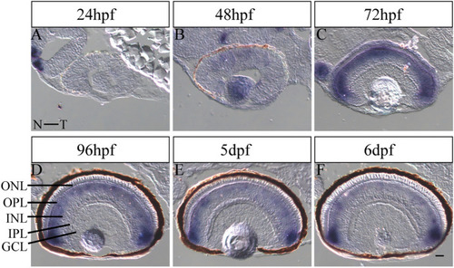
Expression pattern of |
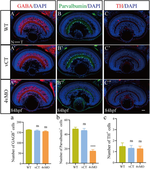
Effects of |
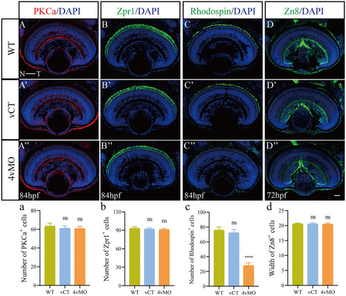
Effects of |
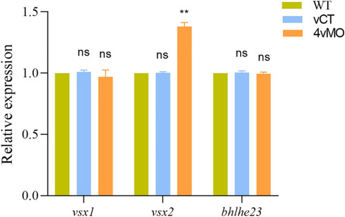
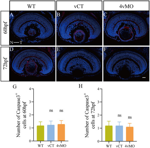
Effects of |
Effects of |
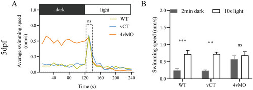
The response of zebrafish larvae to the light stimulus at 5 dpf. ( |