
- Title
-
Genetic evidence for Amh modulation of gonadotropin actions to control gonadal homeostasis and gametogenesis in zebrafish and its noncanonical signalling through Bmpr2a receptor
- Authors
- Zhang, Z., Wu, K., Ren, Z., Ge, W.
- Source
- Full text @ Development
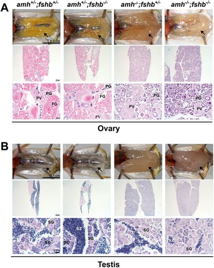
Gonadal histology of amh mutant and double mutant with fshb mutation at 4 mpf. (A) Ovaries of four different genotypes: control (amh+/−;fshb+/−), fshb single mutant (amh+/−;fshb−/−), amh single mutant (amh−/−;fshb+/−) and amh and fshb double mutant (amh−/−;fshb−/−). (B) Testes of the four different genotypes. SC, spermatocytes; SG, spermatogonia; SZ, spermatozoa. Arrows indicate ovaries or testes. PHENOTYPE:
|

Evidence for involvement of gonadotropin signaling in amh deficiency-induced gonadal hypergrowth and dysfunctional gametogenesis. (A) Ovaries of different genotypes at 5 mpf. The amh single mutation (amh−/−;fshr+/−) caused ovarian hypertrophy with accumulation of PG follicles, whereas the fshr single mutant (amh+/−;fshr−/−) showed ovarian hypotrophy with much fewer, underdeveloped PG follicles only. Double mutation of amh and fshr (amh−/−;fshr−/−) completely abolished the phenotype of amh mutant. (B) Testes of different genotypes at 5 mpf. The amh mutation alone (amh−/−;fshr+/−;lhcgr+/−) caused testicular hypertrophy and dysfunctional spermatogenesis with limited meiosis, whereas the loss of gonadotropin signaling in the fshr and lhcgr double mutant (amh+/−;fshr−/−;lhcgr−/−) led to testicular hypotrophy and dysfunctional spermatogenesis, with no meiosis. The loss of all the three genes in the triple knockout (amh−/−;fshr−/−;lhcgr−/−) completely abolished the hypertrophic phenotype of amh mutation. LV, late vitellogenic; SC, spermatocytes; SG, spermatogonia; SZ, spermatozoa. Arrows indicate ovaries or testes. PHENOTYPE:
|

Gonadal hypertrophy in bmpr2a mutant. (A) Ovaries of different genotypes at 5 mpf. The bmpr2a mutation (bmpr2a−/−) caused significant enlargement of the ovary, which contained a large number of PG follicles, with only a few entering PV and vitellogenic growth. (B) Testes of different genotypes at 5 mpf. The bmpr2a mutation also induced hypertrophic growth of the testis, which contained abundant spermatogonia with limited meiosis. (C) Body length, body weight and GSI of different genotypes (n=5; *P<0.05, **P<0.01; two-tailed Student's t-test for unpaired data). (D) Sex ratios in different genotypes. SC, spermatocytes; SG, spermatogonia; ST, spermatids; SZ, spermatozoa. Arrows indicate ovaries or testes. PHENOTYPE:
|

Phenotype analysis of amh and bmpr2a double mutant at 3 mpf. (A) Ovaries of different genotypes. There was a significant accumulation of PG follicles in the amh single mutant (amh−/−;bmpr2a+/−), bmpr2a single mutant (amh−/−;bmpr2a−/−) and amh and bmpr2a double mutant (amh−/−;bmpr2a−/−) in comparison with the control (amh+/−;bmpr2a+/−). (B) Testes of different genotypes at 3 mpf. All three mutants (amh and bmpr2a single mutants and the double mutant) displayed tremendous testicular hypertrophy, with abundant spermatogonia (circled with dotted lines) and without any additive effect. (C) GSI of the different genotypes (n=5; **P<0.01; two-tailed Student's t-test for unpaired data). (D) Viable offspring (3 dpf) from male and female double mutants (amh−/−;bmpr2a−/−). (E) Expression of fshb in the pituitary of male amh and bmpr2a single and double mutants. The groups with different letters indicate that they are statistically significant (P<0.05) [n=4 (amh+/−;bmpr2a+/−), 7 (amh+/−;bmpr2a−/−), 4 (amh−/−;bmpr2a+/−) and 6 (amh−/−;bmpr2a−/−)]. Data are mean±s.e.m. EXPRESSION / LABELING:
PHENOTYPE:
|

Analysis of the phenotype of the bmpr2b mutant. (A) Normal testicular growth and spermatogenesis in male bmpr2b mutant at 3 mpf. (B) Ovarian defects in female bmpr2b mutant at 3 and 5 mpf. Folliculogenesis was arrested at the MV stage in the bmpr2b mutant at both 3 and 5 mpf. (C) Follicle diameters measured on histological section. Two distant sections from each individual were chosen for quantification of PV and vitellogenic follicles with germinal vesicle visible, and three individuals of each genotype (#1-#3) were analyzed at 3 and 5 mpf. Data are mean±s.e.m. (D) Follicular atresia in the ovary of bmpr2b mutants showing abnormal chorion (arrows in bottom panels of d1 and d2) and hypertrophic granulosa cells (asterisks in bottom panel of d2). Staining with DAPI showed the hypertrophic granulosa cells (asterisks in d3) in comparison with normal follicles (d4). Bottom panels in d1 and d2 show higher magnification of the boxed areas above. SC, spermatocytes; SG, spermatogonia; ST, spermatids; SZ, spermatozoa. Arrows indicate ovaries or testes. PHENOTYPE:
|

ZFIN is incorporating published figure images and captions as part of an ongoing project. Figures from some publications have not yet been curated, or are not available for display because of copyright restrictions. PHENOTYPE:
|
|
ZFIN is incorporating published figure images and captions as part of an ongoing project. Figures from some publications have not yet been curated, or are not available for display because of copyright restrictions. |