FIGURE SUMMARY
Title

Functional Conservation of Divergent p63-Bound cis-Regulatory Elements

Authors
Gallardo-Fuentes, L., Santos-Pereira, J.M., Tena, J.J.
Source
Full text @ Front Genet

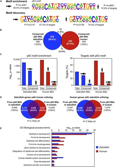
Sequence divergence but functional conservation of p63 binding between zebrafish and human. (A) p63 motif logo found by either motif enrichment (top) or de novo motif discovery (bottom) in p63 ChIP-seq from zebrafish embryos (left) and human keratinocytes (right). Enrichment p-value and percentage of targets with the motif are represented. (B) Venn diagram showing the overlap between p63 binding sites (BSs) in zebrafish and human conserved at the sequence level. (C) p63 motif enrichment (left) and percentage of targets with p63 motif (right) for total and conserved p63 BSs in zebrafish and human. (D) Venn diagrams showing the overlap between zebrafish p63-associated genes with human ortholog (left) or human p63-associated genes with zebrafish ortholog (right) from BSs in both species. P-values for overlap significance using the Hypergeometric test are shown. (E) Gene Ontology (GO) biological process term enrichment for zebrafish and human p63 BSs. Terms related to p63 known functions were selected. Total GO terms are shown in Supplementary Dataset.

Zebrafish and human p63 binding sites (BSs) drive similar expression patterns. (A) Table showing the overlap in expression patterns driven by cis-regulatory element (CRE) pairs from zebrafish and human, as detected in zebrafish transgenic reporter assays. Note that all BS pairs share at least three expression domains in both species. (B) Genome tracks from zebrafish (top) and human (bottom) showing p63 ChIP-seq signal, p63 peaks, ENSEMBL genes, and vertebrate sequence conservation. The selected p63 BSs for transgenic reporter assays, which are located upstream to the myh9a/MYH9 genes, are highlighted in light orange. (C) Transgenic zebrafish embryos at different stages showing the green fluorescent protein (GFP) expression pattern driven by the enhancers highlighted in (B). Tissues expressing GFP are pointed using white arrowheads. s, skin; ysl, yolk syncytial layer; pfb, pectoral fin bud.

Acknowledgments
This image is the copyrighted work of the attributed author or publisher, and ZFIN has permission only to display this image to its users. Additional permissions should be obtained from the applicable author or publisher of the image. Full text @ Front Genet