
- Title
-
Disruption of integrin α4 in zebrafish leads to cephalic hemorrhage during development
- Authors
- Iida, A., Wang, Z., Sehara-Fujisawa, A.
- Source
- Full text @ Genes Genet. Sys.
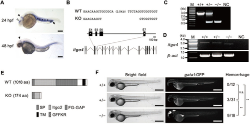
Generation and phenotypic observation of the itgα4 ko108 mutant line. (A) Whole-mount in situ hybridization for itgα4 at 24 and 48 hpf. A 1,001-bp cDNA fragment from the initiation codon was used as the hybridization probe. Strong signals were observed around the eyes (white arrowhead), cerebellum (black arrowheads) and trunk region including the somite, notochord intermediate cell mass (asterisks), and posterior blood island (arrow). Scale bar, 200 μm. (B) Genomic sequence and structure for the itgα4 deletion mutation in zebrafish. The target sequences for the CRISPR/Cas9 were 5’-TCAGAACAAGCTGCCGCA-3’ and 5’-TCAAAGCACTGAGATAGT-3’ (asterisks). (C) PCR-mediated genotyping for the itgα4 mutant. The long (2.6-kb) amplicon means the wild-type allele, and the short (0.6-kb) one is the mutant allele. PCR was carried out using KOD-FX (Toyobo) under the following conditions: 100 s at 94 ℃, followed by 40 cycles of 20 s at 94 ℃, 20 s at 60 ℃ and 120 s at 72 ℃; and 150 s at 72 ℃. Primer sequences are 5’-AACCAGTGGCTGGGTGTGAGTTTG-3’ and 5’-GCGATAAGACTGCATTGTATGTGG-3’. M, size marker. NC, negative control (no template). (D) RT-PCR for itgα4. PCR was carried out using KOD-FX under the following conditions: 100 s at 94 ℃, followed by 35 cycles of 20 s at 94 ℃, 20 s at 60 ℃ and 150 s at 72 ℃; and 210 s at 72 ℃. Primer sequences are [itgα4: 5’-ATGATTTCATCACAACTCACTGGA-3’ and 5’-TTATGATACGCTTTCATGCTTGGG-3’] and [β-actin: 5’-ATGGATGAGGAAATCGCTGCCCTG-3’ and 5’-TTAGAAGCACTTCCTGTGAACGAT-3’]. The longer (> 3.0-kb) amplicon means the wild-type transcript for the functional itgα4 protein. The shorter amplicon (< 3.0 kb) is presumed to correspond to the truncated itgα4 protein. β-actin is an internal control. M, size marker. NC, negative control (no template). (E) Putative structure of the truncated protein synthesized from the mutant allele. The mutant transcript results from a frame-shift mutation and possesses a premature termination codon. SP, signal peptide. TM, transmembrane. (F) Typical images of an embryo for each genotype at 50 hpf. Green fluorescence in Tg (gata1:GFP) ko111 indicates erythrocytes and their progenitor cells (Iida et al., 2018). Accumulation of fluorescent cells means that hemorrhage occurred due to extravascular leakage of circulating erythrocytes. Scale bars, 500 μm. The fractions indicate hemorrhage ratios in each genotype. Chi-squared test was used for statistical analyses. n.s., not significant; **P < 0.01. |

Unillustrated author statements PHENOTYPE:
|