FIGURE SUMMARY
Title

TLR4 signaling drives mesenchymal stromal cells commitment to promote tumor microenvironment transformation in multiple myeloma

Authors
Giallongo, C., Tibullo, D., Camiolo, G., Parrinello, N.L., Romano, A., Puglisi, F., Barbato, A., Conticello, C., Lupo, G., Anfuso, C.D., Lazzarino, G., Li Volti, G., Palumbo, G.A., Di Raimono, F.
Source
Full text @ Cell Death Dis.

SMM- and MM-MSC activated neutrophils in immunosuppressive and pro-angiogenic cells.

a Only SMM- and MM-MSCed-N exhibited suppressive effects compared to N control (isolated from PBMC cultured without MSC). CTRL+ : T lymphocytes incubated only with PHA. b Before incubation with T cells, ed-N were analyzed for the expression of immune modulatory factors. Calculated value of 2−ΔΔCt in control (HC-MSC educated-neutrophils) was 1. c Adding BTZ, LENA or POMA during co-culture of PBMC with MM-MSC, isolated neutrophils did not lose immunosuppressive ability. Ref MM-MSC: MSC isolated from patients with refractory MM. d HMEC were plated on Matrigel in the absence (1, control) or presence of VEGF-A (2, positive control), of SMM-MSCed-N (3), MM-MSCed-N (4), MM-MSCed-N isolated from co-culture with BTZ (5) or LENA (6), refractory MM-MSCed-N (7) or refractory MM-MSCed-N isolated from co-culture with POMA (8). After 5 h, SMM-MSCed-N and refractory MM- and MM-MSCed-N induced tube formation. The pro-angiogenic effect was significantly reduced by the proteasome inhibitor and the immunomodulatory drugs. *p < 0.05; **p < 0.01; ***p < 0.001

PC activated MSC towards an inflammatory phenotype.

a Neutrophils isolated from co-culture with HS-5 or HC-MSC 24 h before pre-treatment with MM cells were able to inhibit T cell proliferation (b) and showed pro-angiogenic capacity in vitro. 1: HMEC control; 2: HMEC in presence of VEGF-A (positive control); 3: plus HC-MSCed-N; 4: plus HC-MSCed-N isolated from co-culture with HC-MSC pre-treated with MM cells. Magnification ×100. c Detection of NF-kB and IRF3 nuclear translocation was performed by incubation respectively with anti-mouse and anti-rabbit monoclonal antibodies followed by secondary antibodies conjugated to FITC (green) or TRITC (red). Counterstaining of cells was performed by using the nuclear dye, DAPI (blue). The photographs result from sequential analysis of the same microscopic field, followed by merging of different images with specific staining. *p < 0.05; **p < 0.01

LPS induced the same immunological alteration of MM-MSC in healthy MSC.

a Neutrophils isolated from co-culture with HC-MSC pre-treated with LPS were able to inhibit T cell proliferation. b After the pre-treatment with LPS, HC-MSC educated-neutrophils showed pro-angiogenic capacity in vitro. 1: HMEC control; 2: HMEC in presence of VEGF-A (positive control); 3: HC-MSCed-N; 4: plus ed-N isolated from co-culture of PBMC with HC-MSC pre-treated with LPS. Magnification ×100. c MyD88 expression was increased in MM-MSC (n = 6) compared to HC-MSC (n = 3). For western blotting analysis, the optical density of the bands was measured using Scion Image software. All showed results represent the means of four independent experiments; error bars denote SD. *p < 0.05; ***p < 0.001

MM-MSC and SMM-MSC enhanced PC engraftment and promoted immune escape mechanisms in zebrafish.

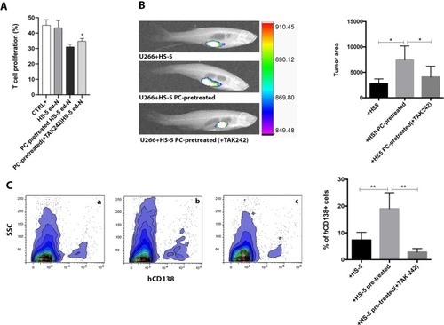
Three animals were engrafted for every combination of PC with SMM-MSC (n = 3), MM-MSC (n = 8) or HC-MSC (n = 4). a Evaluation of tumor xenografts was performed by tomography after 6 days. b Detection of human MM cells using flow cytometry. c Analysis of changes in the Th1/Th2 balance in zebrafish using real time PCR. Results for animals co-injected with PC and MM-MSC are shown (compared to animals injected with PC and HC-MSC); calculated value of 2−ΔΔCt in zebrafish injected with U266 and HC-MSC was 1. All analyses were performed 6 days after cellular mixture injection. *p < 0.05; **p < 0.01; ***p < 0.001

Inhibition of TLR4 signaling in MM-MSC decreased PC engraftment in vivo.

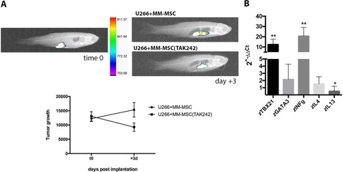
MM-MSC (from five patients) were pre-treated with TAK-242 before co-injection with PC. Three animals were engrafted for every group. a Evaluation of tumor xenograft was performed by tomography after 3 days. Tumor growth within 3 days of engraftment in animals injected with PC + MM-MSC: p < 0.05; tumor reduction within 3 days in animals injected with PC + MM-MSC pretreated with TAK-242 5 μM: p < 0.01. Difference between the two groups: p < 0.001. b Analysis of changes in the Th1/Th2 balance in zebrafish. *p < 0.05; **p < 0.01

Effects of the inhibition of TLR4 during co-culture in vitro of HS-5 with MM cell lines (U266, OPM2, H929) before injection in zebrafish.

a Evaluation of inhibitory capacity of ed-N obtained from coculture of HS-5 pre-treated with MM cells in presence or not of TAK-242. b Evaluation of tumor xenograft was performed by tomography after 3 days. Eight animals were engrafted for every group. (c) hCD138 infiltrate was detected by using flow cytometry. a U266 + HS-5; b U266 + HS-5 pre-treated; c U266 + HS-5 pre-treated (+TAK-242). *p < 0.05; **p < 0.01

Acknowledgments
This image is the copyrighted work of the attributed author or publisher, and ZFIN has permission only to display this image to its users. Additional permissions should be obtained from the applicable author or publisher of the image. Full text @ Cell Death Dis.