
- Title
-
In Vivo Surface Electrocardiography for Adult Zebrafish
- Authors
- Zhao, Y., Yun, M., Nguyen, S.A., Tran, M., Nguyen, T.P.
- Source
- Full text @ J. Vis. Exp.

Contrasting anatomy and ECG of human and zebrafish hearts. In contrast to the human heart with two atria and two ventricles, the zebrafish heart has only one atrium and one ventricle (top row). Abbreviations: RA, right atrium; LA, left atrium; RV, right ventricle; LV: left ventricle. The zebrafish heart shares several common ECG features with the human heart (bottom row). |
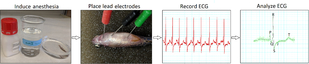
Minimally invasive in vivo ECG recording protocol. A schematic flow chart illustrates four critical action steps in conducting an in vivo ECG interrogation: induce anesthesia, place ECG lead electrodes, record ECG, and analyze the ECG recordings. |
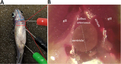
ECG lead placement. Three 29-gauge color-coded stainless steel electrodes are inserted securely into the fish musculature to approximately 1 mm in depth. Placement of the negative (black) electrode and the positive (red) electrode establishes a bipolar lead in the frontal plane, along a left caudal to right cranial orientation. Abbreviation: ref, reference electrode |