FIGURE SUMMARY
Title

Monitoring antiangiogenesis of bevacizumab in zebrafish

Authors
Zhang, J., Gao, B., Zhang, W., Qian, Z., Xiang, Y.
Source
Full text @ Drug Des Devel Ther

Bevacizumab inhibits zebrafish retinal angiogenesis.
(AD) Representative fluorescent images of control or embryos treated with bevacizumab at 4 dpf. In the control image panel, yellow lines indicate the vessel segments. Compared with control, higher magnification of the retinae revealed that collapsed vessels can be observed after bevacizumab treatment. (E) Quantification of the average number of blood vessel segments per eye shows a 11-fold decrease in bevacizumab-treated embryos (n=10) at 4 dpf. *Refers to P<0.0001 by an unpaired Student’s t-test.

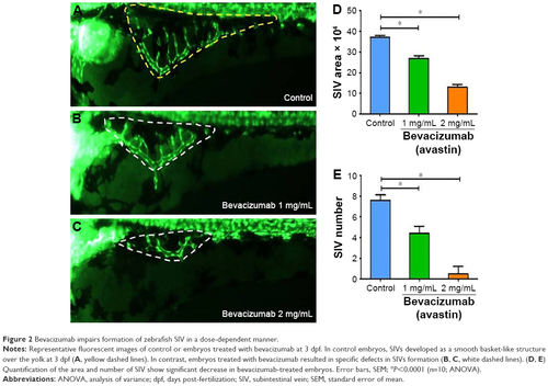
Bevacizumab impairs formation of zebrafish SIV in a dose-dependent manner.
Representative fluorescent images of control or embryos treated with bevacizumab at 3 dpf. In control embryos, SIVs developed as a smooth basket-like structure over the yolk at 3 dpf (A, yellow dashed lines). In contrast, embryos treated with bevacizumab resulted in specific defects in SIVs formation (B, C, white dashed lines). (D, E) Quantification of the area and number of SIV show significant decrease in bevacizumab-treated embryos. Error bars, SEM; *P<0.0001 (n=10; ANOVA).

Bevacizumab causes specific vasculature formation defects in the SIV.
(AD) Representative fluorescent images of zebrafish embryos at 3 dpf treated with PBS (control) or bevacizumab (2 mg/mL). (A, B) In contrast, treatment with bevacizumab at the pectoral fin stage (2.5 dpf) causes specific vasculature formation defects in the SIV (B, white arrow). (CD) Truncal vasculature of the zebrafish shows no appreciable phenotypic differences relative to control.

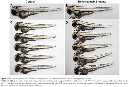
The vascular defect in SIV (subintestinal vein) resulted in further complications such as severe pericardial edema.
(AD) Representative bright field images of zebrafish embroys at 3 days post-fertilization (dpf) treated with PBS (control) or bevacizuamb (2 mg/mL). The vascular defect in SIV resulted in further complications such as severe pericardial edema (B, black arrow) after bevacizumab treatment. Circulation in the SIV is visible in the control fish, but is absent in the bevacizumab-treated zebrafish.

Representative fluorescent images of zebrafish retinal angiogenesis at 4 dpf treated with control (PBS) and bevacizumab (2 mg/mL).
Each row shows 5 examples of the phenotype induced in retina.

Acknowledgments
This image is the copyrighted work of the attributed author or publisher, and ZFIN has permission only to display this image to its users. Additional permissions should be obtained from the applicable author or publisher of the image. Full text @ Drug Des Devel Ther