
- Title
-
Pupil mask diversity for image correction in microscopy
- Authors
- Wilding, D., Pozzi, P., Soloviev, O., Vdovin, G., Verhaegen, M.
- Source
- Full text @ Opt. Express

(a) the evolution of the image acquisition and reconstruction for a plane 75μm inside the zebrafish (b) a ROI at plane 135μm with no mask compared with the first object reconstruction and the last. (c) the wedge positions for images 1–4. (d) the experimental wedge with the pupil size overlayed. (e) the line profile labelled LP in (b). The red arrow shows a feature not present in the original LSFM image that is now clearly resolved. All images are normalised to a 16-bit range minimum to maximum with the colour-scale as shown. |
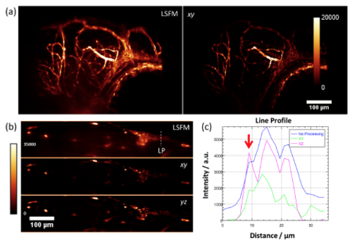
(a) A xy maximum intensity projection of the three-dimensional dataset of the zebrafish larvae head with the standard LSFM (8 datasets) and the diversity processed set plane-by-plane in the xy direction. (b) A slice through the zebrafish in the yz-plane showing the comparison between xy processing and yz processing. (c) A plot of the line profile in (b). All images are normalised to a 16-bit range minimum to maximum with the colour-scale as shown. |