
- Title
-
Expression of meis and hoxa11 in dipnoan and teleost fins provides new insights into the evolution of vertebrate appendages.
- Authors
- Langellotto, F., Fiorentino, M., De Felice, E., Caputi, L., Nittoli, V., Joss, J.M.P., Sordino, P.
- Source
- Full text @ EvoDevo

meis1.1 and hoxa11b expression in zebrafish fins. a–f Dorsal, g, h anal and i–l pelvic fins. a–d, f, h, i–l WISH and e, g Alcian Blue staining followed by WISH. a–c, e, g, h Arrowheads indicate fin mesenchyme cells expressing meis1.1 and hoxa11b. c The white arrow indicates unsegmented radial; c the black arrows indicate WISH staining in dorsal somite cells. a–d, j, l The dashed lines indicate distal limit of endochondrogenic mesoderm below the finfold in a–d dorsal and j, l pelvic fins. e–h The solid lines indicate the boundary between proximal and distal radials in e, f dorsal and g, h anal fins. a–f, i–l Anterior to left, distal to top; g, h anterior to left, distal to bottom. Fish length is a–c 3.6–4.4 mm, d 4.0–5.2 mm, e–h 5.6–6.1 mm, i, k 4.1–5.3 mm, j, l 5.0–6.4 mm. Abbreviations: dr, distal radial; pr, proximal radial. Scale bars are 100 μm (a, b, f, g), 50 μm (c–e, h–l), and 70 μm (k–n) EXPRESSION / LABELING:
|
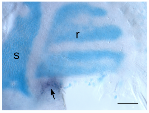
meis1.1 expression and chondrogenesis in zebrafish pectoral fin. Dorsal view of Alcian Blue staining followed by WISH. meis1.1 transcript is restricted to proximal-posterior margin (arrow) during cartilage remodelling. Abbr.: s, scapulocoracoid; r, radial. Scale bar: 100 μm. EXPRESSION / LABELING:
|