
- Title
-
Complementary expression of calcium binding proteins delineates the functional organization of the locomotor network
- Authors
- Berg, E.M., Bertuzzi, M., Ampatzis, K.
- Source
- Full text @ Brain Struct. Funct.
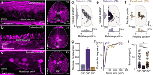
Overview of calretinin (CR), calbindin (CB) and parvalbumin (PV) expression in the adult zebrafish spinal cord. a–c Expression of CBPs in whole mount and transverse sections of the adult zebrafish spinal cord (segment 15). d–f Representative setting positions of the CR, CB and PV positive neurons in spinal hemisegments 15. g Number of CR, CB and PV positive neurons per adult zebrafish spinal hemisegment. h, i Cumulative frequency (h) and average of soma sizes for CR, CB and PV positive neurons. The soma size between CR, CB and PV containing neurons is different (one-way ANOVA: F(2, 645) = 110, p < 0.0001). Data are presented as mean ± SEM; asterisks indicate statistical significance. *p < 0.05; ****p < 0.0001 |

Co-distribution and co-localization of the CBPs positive neurons. a Superimposed positions of the CR, CB and PV positive neurons in the spinal cord. b–d Double immunofluorescent images between the three studied CBPs (CR, CB and PV). Many double expressing neurons observed only between CR and PV (51%; arrowheads in d). e Percentage of co-expression of CR, CB or PV in neurons. Only a small population of PV+ neurons does not express CR (8%; e). Single channel views of the respective framed box |
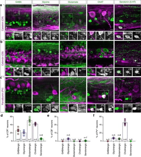
CR, CB and PV expression in neurons with identified neurotransmitter type. a–c Double immunostaining experiments for CR, CB and PV (magenta) with markers that label the GABAergic, glycinergic, glutamatergic, cholinergic and serotonergic spinal neurons (green) in whole mount adult zebrafish spinal preparations. Enlarged and single channel views are given for areas indicated by framed boxes. Arrowheads indicate double-labeled cells. d–f Quantification of the percentage of CR, CB and PV positive neurons that localized in neurons with a specific neurotransmitter phenotype |

Zebrafish axial motoneurons are immunoreactive to CR and PV. a Representative images of the double-labeled motoneurons with CR and PV. b Quantification of the percentage of the motoneurons that express both CR and PV. c Whole mount images showing the expression of CR and PV in distinct motoneuron pools (slow, intermediate and fast). Single channel views of the respective images shown for better visualization of CR or PV positive cells. Arrowheads indicate the double-labeled cells. d Color coded quantification of the CR and PV expression in distinct populations of neurons. The vast majority of motoneurons contain PV. CR expression is localized mainly in the motoneurons of the fast module |
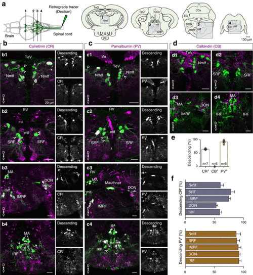
Distribution pattern of CR, CB and PV expression in reticulospinal neurons. a Schematic representation of the methodology used to reveal the brain neurons projecting to the spinal cord. The analyzed brain areas are indicated by boxes in coronal sections 1–4. b–d Microphotographs from coronal brain sections, showing the distribution of the brain neurons that project to the spinal cord (green) and the CR, CB and PV expression (magenta). Single channel views of the respective images shown for better visualization of the double-labeled neurons. Arrowheads indicate the double-labeled cells. e Percentage of the reticulospinal neurons that express CR, CB or PV, respectively. f Bar graph showing the percentage of brain to spinal cord projecting neurons in several brain areas that express CR or PV |
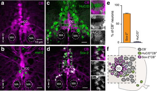
Identity of the CB positive neurons. a Distribution pattern of CB+ expression around the central canal in the adult zebrafish spinal cord. b None of the CB expressing cells (magenta) co-localized with the early neuronal differentiated marker mef-2 (green). c–d A small number (2.32%) of the differentiated and mature neurons (HuC/D+, green) contain also CB (magenta). In addition, the vast majority (78.13%) of CB containing cells (magenta) is progenitor cells / stem cells (Sox-2+, green). Single channel views shown for areas are indicated by framed rectangles. Arrowheads indicate double labeled cells. e Quantification of CB positive cells that co-express Sox-2+ or HuC/D+. f Schematic representation of the central canal area in the adult zebrafish spinal cord showing that most of the CB+ cells are progenitors / stem cells |