
- Title
-
Impaired AMPA receptor trafficking by a double knockout of zebrafish olfactomedin1a/b
- Authors
- Nakaya, N., Sultana, A., Tomarev, S.I.
- Source
- Full text @ J. Neurochem.

ZFIN is incorporating published figure images and captions as part of an ongoing project. Figures from some publications have not yet been curated, or are not available for display because of copyright restrictions. |
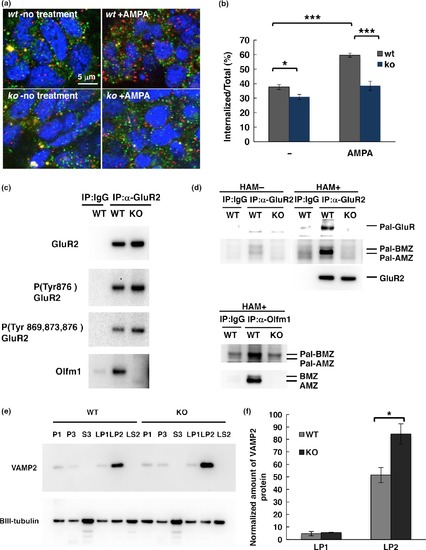
Changes in the localization of GluR2 and VAMP2 in olfm1 null retina and brain. (a) The internalization of GluR2 in olfm1 null and wt larval retinal cells in culture. Retina from 32 hpf larvae were dissected and cultured in vitro for 5 days. External GluR2s were labeled with antibodies against the N‐terminal part of GluR2 and allowed to be internalized for 15 min with or without 100 μM α‐amino‐3‐hydroxy‐5‐methylisoxazole‐4‐propionate in the culture. The external GluR2 was detected using Alexa488‐secondary antibody. The internalized GluR2 was detected using Alexa555‐secondary antibody after permeabilization of the cell membrane. Scale bar, 5 μm. (b) The number of green and red puncta was counted and the ratio of the internal and the total labeled GluR2 was calculated. N = 6–8 individual cultures. (c) The levels of GluR2 and phosphorylated GluR2 were slightly increased in synaptosomes isolated from olfm1 null brain compared with wt brain. (d) Reduced palmitoylation of GluR2 precipitated by anti‐GluR2 antibody from synaptosomes isolated from olfm1 null brain compared with wt samples (upper panel). Note that olfm1 co‐immunoprecipitated with GluR2 was also palmitoylated, indicating that Olfm1 may be associated with membranes through its lipid modification as well as its binding to other membrane‐associated proteins (lower panel). HAM‐untreated precipitates were used as a negative control and they show only weak signals for both GluR2 and Olfm1 (e) The level of VAMP2 was increased only in the synaptosomal LP2 fraction (synaptic vesicles) but not in LP2 (synaptosomal membrane). (f) Quantification of three independent experiments as in (f). *p < 0.05,***p < 0.001. |
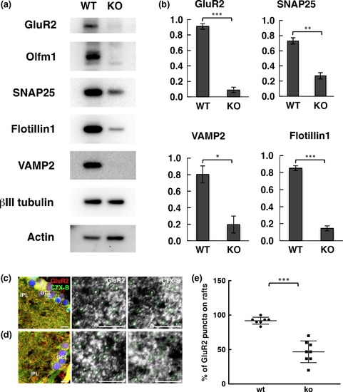
Characterization of the microdomain fraction from wt and olfm1 null brain. (a) Changes in the protein levels detected in the microdomain fraction from wt and olfm1 null adult brain. (b) Quantification of the results of three independent experiments as in (a). (c, d) GluR2 localization in CTX‐B‐labeled lipid rafts in the IPL of adult wt (c) and olfm1 null (d) zebrafish retina. The black/white images in the middle and right rows represent enlarged images in each channel from the red squared areas in the left row. Note that all green‐circled GluR2‐positive puncta are localized in CTX‐B‐bound lipid rafts in wt retina, while some of GluR2 puncta in olfm1 null are not in lipid rafts. Scale bar, 5 μm. (e) Quantification of the results of seven wt and eight olfm1 null independent eye sections as in (c and d). *p < 0.05, **p < 0.01, ***p < 0.001. |