
- Title
-
The E-cadherin/AmotL2 complex organizes actin filaments required for epithelial hexagonal packing and blastocyst hatching
- Authors
- Hildebrand, S., Hultin, S., Subramani, A., Petropoulos, S., Zhang, Y., Cao, X., Mpindi, J., Kalloniemi, O., Johansson, S., Majumdar, A., Lanner, F., Holmgren, L.
- Source
- Full text @ Sci. Rep.
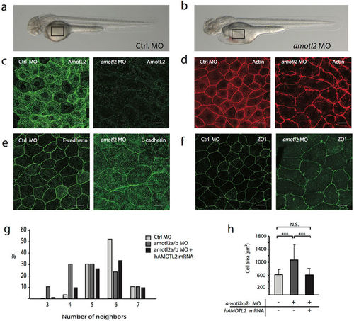
AmotL2 controls actin organization and epithelial cell geometry in zebrafish skin. (a) Bright-field image of a zebrafish embryo 48 hours post fertilization (hpf). (b) Bright-field image of a zebrafish amotl2 morphant. The zebrafish development is apparently normal except for the pericardial edema due to vascular defects as described29. The black square indicates the area of the skin that is visualized in (c–f). Scale bars = 25 µm. (c–f). Confocal microscopy images of whole mount immunostainings of zebrafish skin. (c) Shows the localization of amotL2 in the control fish as indicated and knock-down efficiency after injection of amotl2 MO. (d) Visualization of actin filaments using phalloidin staining. (e) and (f) E-cadherin as adhesion junction marker and ZO1 as tight junction marker. Scale bars = 25 µm, Data are derived from three independent experiments. (g) Quantification of number of neighboring skin cells in Ctrl MO, amotl2 MO and amotl2 MO + p100 mRNA (rescue experiment) zebrafish embryo skin. The control cells are mostly pentagonal and hexagonal, while cells with three or four corners were observed to a higher extent in the amotl2 MO embryos. The phenotype could be rescued by co-injection of a human AMOTL2 p100 mRNA. Differences in distributions between the three populations were evaluated by two-sample Kolmogorov-Smirnov test. Ctrl vs. amotl2 MO, p-value = 2.07 × 10⁻⁵; amotl2 MO vs. amotl2 MO + p100 mRNA, p-value = 0.00029; ctrl vs. amotl2 MO + p100 mRNA p-value = 0.024. N(Ctrl) = 241 cells, n(amotl2 MO) = 228 cells, n(amotl2 MO + p100 mRNA) = 373 cells. Data are derived from three independent experiments. (h) Bar diagrams summarizing the differences in average cell area size of Ctrl MO, amotl2 MO and amotl2 MO + p100 mRNA (rescue experiment) zebrafish embryo skin. Ctrl vs. amotl2 MO, p-value = 4.35 × 10⁻²⁷; amotl2 MO vs. amotl2 MO + p100 mRNA, p-value = 1.75 × 10⁻³⁴; ctrl vs. amotl2 MO + p100 mRNA, p-value = 0.64. N(ctrl) = 144 cells, n(amotl2 MO) = 216 cells, n (amotl2 MO + p100 mRNA) = 227 cells. Data are derived out of three independent experiments. |
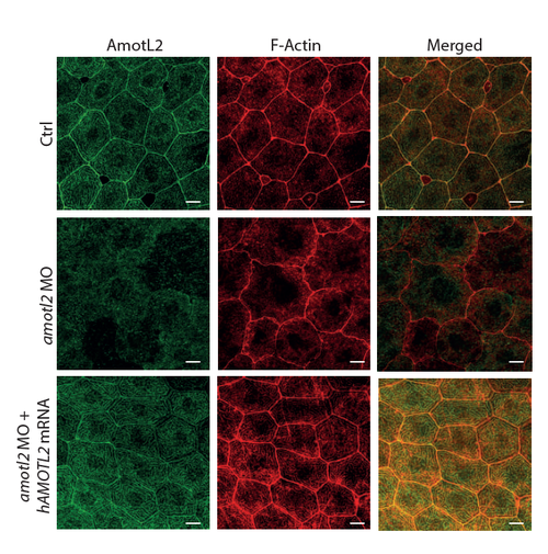
AmotL2 controls actin organization and epithelial cell geometry in zebrafish skin. Filamentous F-actin structures were lost in amotl2 MO epidermis, while junctional actin still seemed somewhat intact. Note the altered cell area and morphology in the skin of the amotl2 MO embryo. The observed phenotype could be rescued by re-expression of human AMOTL2 mRNA. Scale bars=10 μm. Data are derived from three independent experiments. PHENOTYPE:
|