
- Title
-
Somite-Derived Retinoic Acid Regulates Zebrafish Hematopoietic Stem Cell Formation
- Authors
- Pillay, L.M., Mackowetzky, K.J., Widen, S.A., Waskiewicz, A.J.
- Source
- Full text @ PLoS One
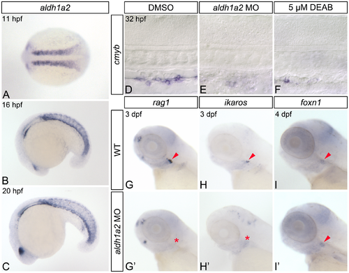
RA-deficient embryos demonstrate impaired HSC formation. (A-C) In situ hybridization analyses of aldh1a2 gene expression in wild type (WT) embryos. (A) Expression within the somites at 11 hpf, shown in dorsal view with anterior to the left. Somitic expression persists in 16 hpf (B) and 20 hpf (C) embryos, shown in lateral view with anterior to the left. (D-F) Representative flat-mounted embryos following in situ hybridization analysis of cmyb gene expression at 32 hpf. Lateral view of gene expression in the dorsal aorta region of the trunk is shown with anterior to the left. Compared to DMSO-treated controls (D) aldh1a2-morphants (E), and 5 μM DEAB-treated embryos (F) exhibit nearly abolished cmyb expression. (G-H') In situ hybridization analyses of common lymphoid progenitor gene expression in 3 dpf embryos. Lateral view of gene expression in the head is shown with anterior to the left. Arrowheads and asterisks indicate thymus. Compared to WT embryos (G, H), aldh1a2-morphants exhibit nearly abolished thymic rag1 (G') and ikaros (H') expression. (I, I') Representative embryos following in situ hybridization analysis of foxn1 thymic epithelial cell gene expression in 4 dpf embryos. Lateral view of gene expression in the head is shown with anterior to the left. Arrowheads indicate thymus. WT embryos (I) and aldh1a2-morphants (I') exhibit similar thymic foxn1 expression levels. |
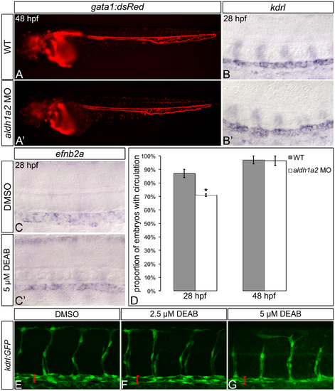
RA-deficient embryos exhibit relatively normal gross embryonic vasculogenesis. (A, A') Lateral view of live 48 hpf Tg(gata1:DsRed)sd2Tg embryos with anterior to the left. Compared to wild type (WT) embryos (A), aldh1a2-morphants (A') display visible circulating blood cells, and an intact dorsal aorta and posterior cardinal vein. (B-C') Representative embryos following in situ hybridization analysis of kdrl vasculature marker gene expression (B, B') or efnb2a arterial marker gene expression (C, C') in 28 hpf embryos. Lateral view of gene expression in the dorsal aorta region of the trunk is shown in flat-mount embryos, with anterior to the left. Compared to WT embryos (B), aldh1a2-morphants (B') exhibit normal dorsal aorta kdrl gene expression. Compared to DMSO-treated controls (C), embryos treated with 5 μM DEAB (C') exhibit normal levels, but a reduced domain of dorsal aorta efnb2a gene expression. (D) Graph demonstrating the mean proportion of WT or aldh1a2-morphant embryos with intact circulation at 28 hpf and 48 hpf. Error bars represent standard error. *Indicates statistically significant difference compared to WT (P = 0.0196). See text for statistical tests. (E-G) Lateral view of dorsal aorta region of the trunk is shown in representative flat-mount Tg(kdrl:GFP)la116Tg 28 hpf embryos, with anterior to the left. Brackets indicate dorsal aorta. Compared to DMSO-treated controls (E), 2.5 μM DEAB-treated embryos (F) exhibit normal dorsal aorta morphology, while 5 μM DEAB-treated embryos (G) exhibit thinning of the dorsal aorta. |
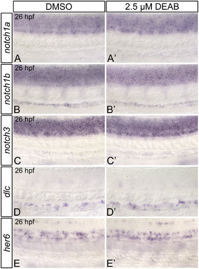
RA-deficient embryos demonstrate normal dorsal aorta notch and Notch1-target gene expression. Representative flat-mount 26 hpf embryos following in situ hybridization analyses. Lateral view of gene expression in the dorsal aorta region of the trunk is shown with anterior to the left. Compared to DMSO-treated controls (A, B), embryos treated with 2.5 μM DEAB exhibit normal notch1a (A'), and notch1b (B') gene expression within the trunk and dorsal aorta. notch3 is expressed at normal levels in the dorsal aorta (C, C'), but is mildly upregulated in the somites of 2.5 μM DEAB-treated versus DMSO-treated control embryos (data not shown). Compared to DMSO-treated controls (D, E) embryos treated with 2.5 μM DEAB exhibit normal gene expression levels of the Notch1-signaling pathway transcriptional targets dlc (D') and her6 (E'). |
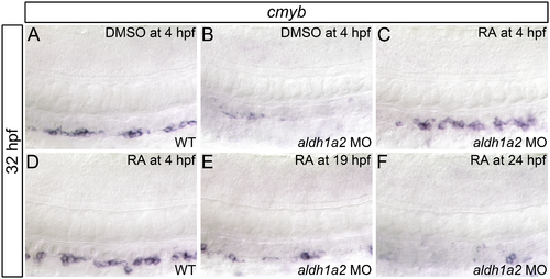
RA is required prior to 19 hpf for HSC formation. Shown are representative flat-mount embryos following in situ hybridization analyses of cmyb gene expression in wild type (WT; A, D) or aldh1a2-morphant (B, C, E, F) 32 hpf embryos treated with DMSO (A, B) or 1 nM RA (C-F) at indicated time points. Lateral view of gene expression in the dorsal aorta region of the trunk is shown with anterior to the left. Compared to WT embryos (A), embryos treated with 1 nM RA (D) exhibit normal cmyb expression (two-tailed P = 1.000). aldh1a2-morphants (B) exhibit nearly abolished cmyb expression compared to WT embryos (two-tailed P = 0.01). cmyb expression is significantly restored in aldh1a2-morphant embryos treated with 1 nM RA at 4 hpf (C; two-tailed P = 1.000 compared to WT). cmyb expression is not significantly restored in aldh1a2-morphants treated with 1 nM RA at 19 hpf (E; two-tailed P < 0.0005) or 24 hpf (F; two-tailed P < 0.0005). See text for statistical tests. |

RA does not regulate the somitic expression of Wnt16-Notch3 signaling pathway components. Shown are representative 17 hpf embryos following in situ hybridization analyses (A-J'). Lateral view (A-J) or dorsal view (A'-J') of gene expression is shown with anterior oriented to the left. A'-J' represent different views of the embryos shown in A-J. Compared to DMSO-treated controls (A, A', C, C', E, E', G, G'), DEAB-treated embryos exhibit normal somitic expression levels of wnt16 (B, B'), and dlc (D, D’), mildly increased dld expression (F, F'), and increased notch3 somitic gene expression (H, H'). DEAB-treated embryos also exhibit normal expression levels of the Notch3 signaling pathway transcriptional target her9 (J, J'), when compared to DMSO-treated controls (I, I'). (K) Quantitative real-time PCR analysis of her9 expression in 17 hpf DMSO-treated controls and embryos treated with 5 μM DEAB. Shown is the relative quantity of her9 expression. Samples were normalized to ef1a and DMSO-treated was set to 1. Error bars indicate standard error of the mean. *Indicates the difference compared to control is significant by Student t test, P = 0.0198. |

RA-deficient embryos exhibit abnormal jam1a and jam2a expression. Representative flat-mount 17 hpf embryos following in situ hybridization analyses. Dorsal view of gene expression is shown with anterior to the left. Compared to DMSO-treated controls (A), embryos treated with 5 μM DEAB (A’) exhibit wild type levels of jam1a expression, and extreme lateral positioning of the anterior-most domains of jam1a expression (double-headed arrows). Compared to DMSO-treated controls (B), embryos treated with 5 μM DEAB (B’) display strongly increased somitic jam2a expression, and lateral expansion of the jam2a expression domain. (C) Graph demonstrating length of the domain of jam2a expression along the medio-lateral axis, divided by length of the domain of expression along the anterior-posterior axis of the eighth jam2a-expressing somite on the right side of the embryo (see asterisk in B, B’). Error bars represent standard error *Indicates statistically significant difference in ratio compared to DMSO-treated controls (P < 0.0001). See text for statistical tests. |
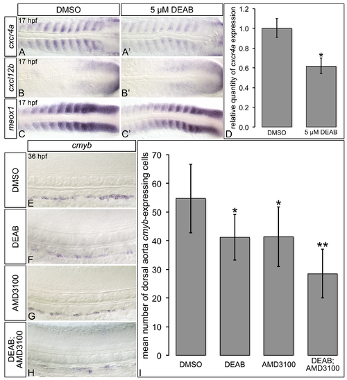
RA-deficient embryos exhibit altered Cxcl12b chemokine signaling pathway component gene expression. (A-C’) Representative flat-mount 17 hpf embryos following in situ hybridization analyses. Dorsal of gene expression is shown with anterior to the left. Compared to DMSO-treated controls (A), embryos treated with 5 μM DEAB (A’) exhibit strongly reduced somitic cxcr4a gene expression, and narrowing of the cxcr4a expression domain within each somite. Compared to DMSO-treated controls (B, C), embryos treated with 5 μM DEAB exhibit subtly increased levels of somitic cxcl12b expression (B’), and subtly decreased levels of somitic meox1 expression (C’). (D) Quantitative real-time PCR analysis of cxcr4a expression in 17 hpf DMSO-treated controls and embryos treated with 5 μM DEAB. Shown is the relative quantity of cxcr4a expression. Samples were normalized to ef1a and DMSO-treated was set to 1. Error bars indicate standard error of the mean. *Indicates the difference compared to control is significant by Student t test, P < 0.0382. (E-H) Representative flat-mount 36 hpf embryos following in situ hybridization analyses of cmyb gene expression. Lateral view of gene expression in the dorsal aorta region of the trunk is shown with anterior to the left. Compared to DMSO-treated controls (E), embryos treated with 1 μM DEAB (F) or 10 μM AMD3100 (G) exhibit a small reduction cmyb-expressing cell numbers. Embryos treated with both 1 μM DEAB and 10 μM AMD310 (H) exhibit a more severe reduction in cmyb-expressing cell numbers. (I) Graph demonstrating the mean number of dorsal aorta cmyb-expressing cells in DMSO-treated controls, embryos treated with 1 μM DEAB, 10 μM AMD3100, or both 1 μM DEAB and 10 μM AMD310. Error bars represent standard error. *Indicates statistically significant difference compared to control (P ≤ 0.0144). **Indicates statistically significant difference compared to 1 μM DEAB, and 10 μM AMD3100 (P ≤ 0.0028). See text for statistical tests. |

28 hpf RA-deficient embryos exhibit increased Cxcl12b chemokine signaling pathway component gene expression. (A-C') Representative flat-mount 28 hpf embryos following in situ hybridization analyses. Lateral view of gene expression in the dorsal aorta region of the trunk is shown with anterior to the left. Compared to DMSO-treated controls (A, B, C), embryos treated with 2.5 μM DEAB exhibit strongly increased levels of dorsal aorta cxcr4a (A'), and cxcl12b (B') gene expression, and upregulated somitic meox1 expression (C'). |