
- Title
-
Antigen Uptake during Different Life Stages of Zebrafish (Danio rerio) Using a GFP-Tagged Yersinia ruckeri
- Authors
- Korbut, R., Mehrdana, F., Kania, P.W., Larsen, M.H., Frees, D., Dalsgaard, I., Jørgensen, L.V.
- Source
- Full text @ PLoS One
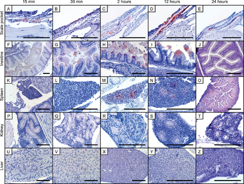
Immunohistochemical images of antigen uptake in adult zebrafish tissues following bath in a Y. ruckeri bacterin. Antigen staining is visible in scale pockets (A-E) and in the intestines from 15 min to 24 hpb (F-J). Two to 24 hpb the Yr bacterin is present in the spleen (M-O) and 24 hpb staining of the bacterin can be observed in the kidney (T) and in the liver (Z). All scale bars are 100 μm long. |

Immunohistochemical visualisation of Y. ruckeri bacterin in the gills of adult zebrafish following bath. Staining of a Y. ruckeri bacterin is observed in the gills of an adult zebrafish 15 min (A) and 30 min (B) following bath. The scale bars are 100 μm long. |

Immunohistochemical images of antigen uptake in juvenile zebrafish tissues following bath in a Y. ruckeri bacterin. Extensive bacterin uptake is seen in the intestine of juvenile zebrafish 30 min post bath (pb) (A). At the basis of the sensory hairs in the nares staining is observed two hpb (B) and the bacterin is subsequently present in the spleen 12 hpb (C). All scale bars are 100 μm long. |
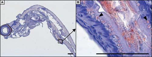
Immunohistochemical staining of antigen uptake in 20 dpf zebrafish larvae 24 h post bath with a Y. ruckeri bacterin. A whole larvae is shown (A), where specialised enterocytes of the mid-intestine has taken up the bacterin (B). The scale bars are 100 μm long. |
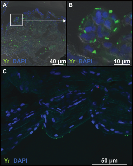
Immunohistochemical staining of the gills and skin of 20 dpf zebrafish larvae following bath with a Y. ruckeri bacterin. Fifteen min post bath the bacterin is present in the gills of zebrafish larvae (A and B) and also on the skin of the fish (C). Bacterin staining is green and nuclei staining is blue (DAPI). |

Images of antigen uptake in a transparent zebrafish following bath in a GFP-tagged Y. ruckeri bacterin. (A) The transparent tra:nac mutant, which was used for the investigations. (B) GFP-tagged Y. ruckeri bacterin on the skin of adults at one min post bath (pb). Bacterin was detected in scale pockets following 15 min (C) and 30 min pb (D). The GFP-tagged bacterin was observed 30 min pb in the intestine and in the oesophagus, which can be realised behind the gills (E). Presence of the bacterin was furthermore detected on the skin and in scale pockets following 30 min (F), 2 h (G) and 12 hpb (H). |
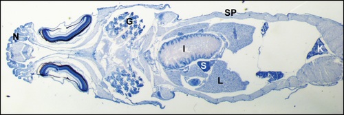
Section of a zebrafish showing organs and tissues relevant for the present study. G is gills, I is intestine, K is kidney, L is liver, N is nose, S is spleen, SP is scale pocket or skin. |
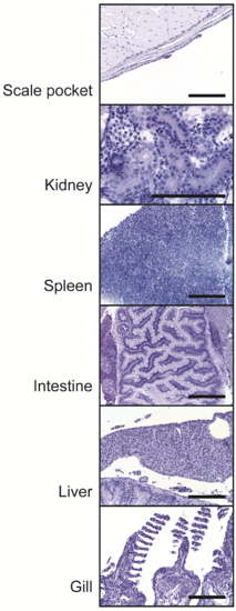
Immunohistochemical staining of adult zebrafish tissues following a sham bath. Sections were stained with an anti-Y. ruckeri antibody and the absence of colour reactions are shown for the scale pocket, the kidney, the spleen, the intestine, the liver and the gill. The scale bars are 100 μm long. |