
Image
|
Figure Caption
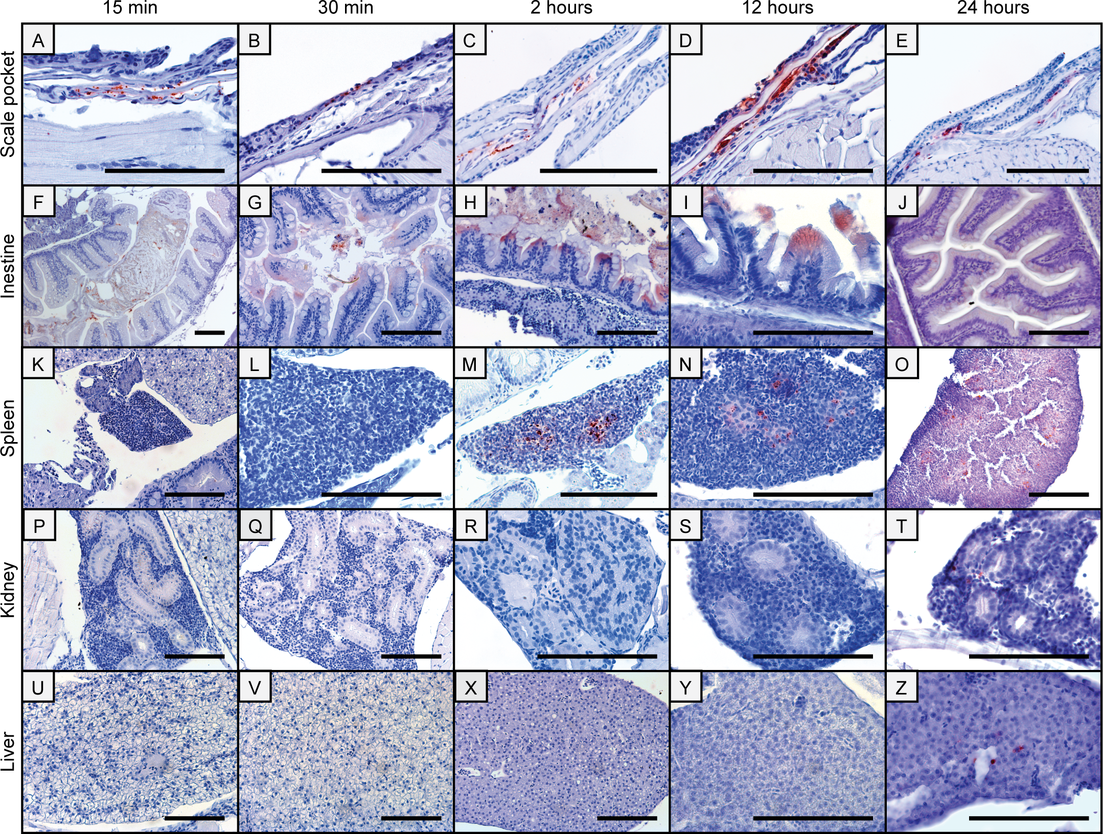
Fig. 1
Immunohistochemical images of antigen uptake in adult zebrafish tissues following bath in a Y. ruckeri bacterin.
Antigen staining is visible in scale pockets (A-E) and in the intestines from 15 min to 24 hpb (F-J). Two to 24 hpb the Yr bacterin is present in the spleen (M-O) and 24 hpb staining of the bacterin can be observed in the kidney (T) and in the liver (Z). All scale bars are 100 μm long.
Acknowledgments
This image is the copyrighted work of the attributed author or publisher, and
ZFIN has permission only to display this image to its users.
Additional permissions should be obtained from the applicable author or publisher of the image.
Full text @ PLoS One