
- Title
-
Role of Active Contraction and Tropomodulins in Regulating Actin Filament Length and Sarcomere Structure in Developing Zebrafish Skeletal Muscle
- Authors
- Mazelet, L., Parker, M.O., Li, M., Arner, A., Ashworth, R.
- Source
- Full text @ Front. Physiol.
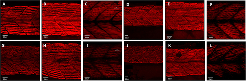
Disruption of myofibril organization during paralysis is reversed by restoring movement in developing skeletal muscle. Control embryos (Embryo Medium alone) and treated embryos (Embryo Medium with Tricaine) were incubated for 7 h starting at 17 hpf. At 24 hpf embryos were removed and control (A) and treated embryos (G) were fixed and stained immediately or put into recovery (Embryo Medium without Tricaine) and (B) control and (H) recovered embryos fixed and stained at 42 hpf. Some embryos were kept in recovery from 24 hpf up to 5 dpf before fixation and staining of both control (C) and recovered larvae (I). Relaxed immotile mutants were fixed and stained at 24 hpf (J), at 42 hpf (K), and 5 dpf (L) as well as motile control siblings at 24 hpf (D), 42 hpf (E) and 5 dpf (F). Anti-myosin antibody (F59) in (A,B,D,E,G,H,J,K), and Rhodamine phalloidin actin labeling in (C,I,F,L) reveals slow muscle fibers. For consistency, the somites imaged were taken at the level where the yolk sac and the yolk sac extension join. Scale bars corresponds to 20 μm. |

ZFIN is incorporating published figure images and captions as part of an ongoing project. Figures from some publications have not yet been curated, or are not available for display because of copyright restrictions. PHENOTYPE:
|
|
ZFIN is incorporating published figure images and captions as part of an ongoing project. Figures from some publications have not yet been curated, or are not available for display because of copyright restrictions. PHENOTYPE:
|
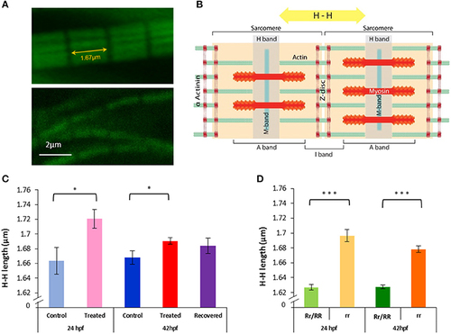
Paralysis leads to actin lengthening in both relaxed mutant and Tricaine treated embryos, subsequent movement restoration in recovered embryos leads to actin length complete rescue. (A,B) H-H measurement. Confocal images of actin filaments stained with phalloidin (1/40) were taken (upper panel 24 hpf control embryo and lower panel 24 hpf Tricaine treated embryo) and measurements between H bands, across Z discs and sarcomeres, were made. (C) 17–24 hpf paralysis caused a significant lengthening of actin filaments H-H at 24 hpf (*p < 0.05) (n = 20 in 5 controls, n = 15 in 3 treated embryos, t-test). At 42 hpf, recovered embryos (17–24 hpf paralysis) showed a full recovery (n = 50 in 10 control embryos, n = 40 in 8 recovered embryos, unpaired t-test), in contrast with treated embryos (17–42 hpf paralysis) which did not recover (n = 50 in 10 control embryos, n = 135 in 27 treated,*p < 0.05). (D) Immotile relaxed mutants, rr, showed a very significant lengthening of actin filaments H-H at 24 hpf (***p < 0.001) (n = 115 in 23 immotile relaxed mutants rr, n = 120 in 24 motile control siblings Rr/RR and at 42hpf (***p < 0.001) (n = 105 in 21 immotile relaxed mutants rr, n = 135 in 27 motile control siblings Rr/RR). Interestingly, in contrast with the Tricaine treated immotile embryos, the immotile relaxed mutant (rr) actin is still very significantly longer than the motile control siblings (Rr/RR) actin at 42 hpf. It is noticeable that in the complete absence of movement up to 42 hpf actin filaments showed a significant shortening between 24 and 42 hpf in between both treated and control embryos and immotile relaxed mutants and motile control siblings, respectively (n = 15 in 3 24 hpf treated, n = 135 in 27 42 hpf treated embryos, *p < 0.05 unpaired t-test; and n = 115 in 23 24 hpf immotile relaxed mutants rr, n = 105 in 21 42 hpf immotile relaxed mutants rr, *p < 0.05 unpaired t-test). |
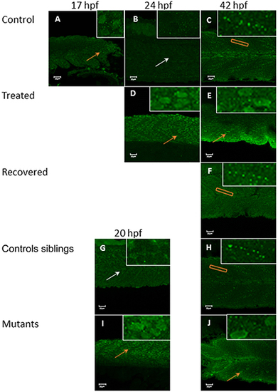
Tropomodulin 1 localization within the fast skeletal muscle from 17 hpf up to 42 hpf in immotile and control embryos. In control embryos (A–C) Tmod1 is initially located in the nucleus of the fast muscle cell at 17 hpf (orange arrow, inserts) (A) and then migrate into the skeletal muscle cell cytosol at 24 hpf (empty nuclei, white arrow, insert) (B) where it stays and get organized linearly by 42 hpf (orange rectangle, insert) (C). Tricaine treated immotile embryos (D,E) display a nuclear localization of Tmod1 at both 24 hpf (D) and 42 hpf (E) (orange arrows, inserts). Recovered motile embryos (F) display at 42 hpf a similar cytosolic alignment of Tmod1 as control embryos (orange rectangle, insert). Controls siblings motile embryos (G,H) display a cytosolic pattern of Tmod1 from 20 hpf (G) (empty nuclei, white arrow, inserts) which is linearized by 42 hpf (H) (orange box, insert) as control and recovered embryos. Immotile relaxed mutants embryos (I,J) display a similar nuclear localization of Tmod1 at both 20 hpf (I) and 42 hpf (J) as Tricaine treated embryos (orange arrows, inserts). Right hand corner inserts shown at a magnification of X5 compared to main image. |
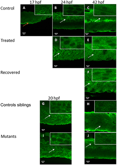
Tropomodulin 4 localization within the fast muscle cell from 17 hpf up to 42 hpf in immotile and control embryos. In control embryos (A–C) Tmod4 is initially located in the nucleus of the fast muscle cell at 17 hpf (orange arrow, insert) (A) and then migrate into the fast muscle cell cytosol at 24 hpf (B) where it stays up to 42 hpf (empty nuclei white arrows, inserts) (C). Tricaine treated immotile embryos (D,E) display a cytosolic localization of Tmod4 at both 24 hpf (D) and 42 hpf (E) (empty nuclei, white arrows, inserts). Recovered motile embryos at 42 hpf (F) display a similar cytosolic localization of Tmod4 as control and treated embryos (empty nuclei, white arrows, inserts). Controls siblings motile embryos (G,H) display a cytosolic localization of Tmod4 from 20 hpf (G) which is maintained up to 42 hpf (H) (empty nuclei, white arrows, inserts). Immotile relaxed mutants embryos (I,J) display a similar cytosolic localization of Tmod4 at both 20 hpf (I) and 42 hpf (J) as control siblings (empty nuclei, white arrows, inserts). Right hand corner inserts shown at a magnification of X5 compared to main image. |