
|
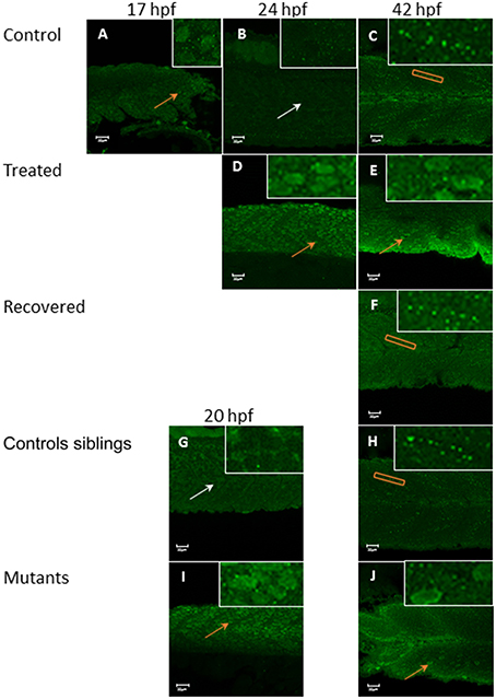
Fig. 7
Tropomodulin 1 localization within the fast skeletal muscle from 17 hpf up to 42 hpf in immotile and control embryos. In control embryos (A–C) Tmod1 is initially located in the nucleus of the fast muscle cell at 17 hpf (orange arrow, inserts) (A) and then migrate into the skeletal muscle cell cytosol at 24 hpf (empty nuclei, white arrow, insert) (B) where it stays and get organized linearly by 42 hpf (orange rectangle, insert) (C). Tricaine treated immotile embryos (D,E) display a nuclear localization of Tmod1 at both 24 hpf (D) and 42 hpf (E) (orange arrows, inserts). Recovered motile embryos (F) display at 42 hpf a similar cytosolic alignment of Tmod1 as control embryos (orange rectangle, insert). Controls siblings motile embryos (G,H) display a cytosolic pattern of Tmod1 from 20 hpf (G) (empty nuclei, white arrow, inserts) which is linearized by 42 hpf (H) (orange box, insert) as control and recovered embryos. Immotile relaxed mutants embryos (I,J) display a similar nuclear localization of Tmod1 at both 20 hpf (I) and 42 hpf (J) as Tricaine treated embryos (orange arrows, inserts). Right hand corner inserts shown at a magnification of X5 compared to main image.