
- Title
-
DNA Damage Response in Proliferating Müller Glia in the Mammalian Retina
- Authors
- Nomura-Komoike, K., Saitoh, F., Komoike, Y., Fujieda, H.
- Source
- Full text @ Invest. Ophthalmol. Vis. Sci.

DNA damage response is absent in zebrafish Müller glia and rat retinal progenitors. (A) Double immunofluorescence for γH2AX/BrdU or p53/BrdU in the zebrafish retina at day 8 after MNU treatment. BrdU+ neurogenic clusters are negative for both γH2AX and p53 (arrows). ONL, outer nuclear layer; INL, inner nuclear layer; GCL, ganglion cell layer. Scale bar: 20 µm. (B) Double immunofluorescence for γH2AX/MCM6 or p53/MCM6 in the rat retina at postnatal day 3. MCM6+ retinal progenitors are negative for both γH2AX and p53. NBL, neuroblastic layer; GCL, ganglion cell layer. Scale bar: 20 µm. EXPRESSION / LABELING:
|
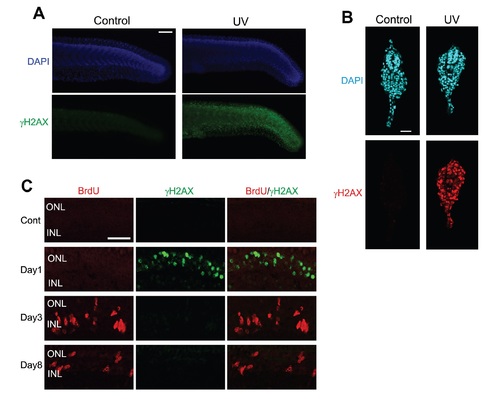
γH2AX immunoreactivity in UV-irradiated zebrafish tissue or zebrafish photoreceptors after MNU treatment. A. Whole-mount immunofluorescence for γH2AX in UV -irradiated zebrafish embryos. Zebrafish embryos at 24 h post fertilization were irradiated to 2.43 J/cm2 ofUV-C by using UVGL-58 handheld UV lamp (UVP, Upland, CA, USA). Embryos were then cultured in a dark incubation chamber for 30 min at 28.5°C and fIXed with 4% paraforrn-aldehyde for 1 h at 4°C. Whole-mount immunofluorescence was as previously described by Komoike and Matsuoka (2013). Scale bar = 100 µm. B. Immunofluorescence for γH2AX in UV -irradiated zebrafish embryos using cryostat sections. Scale bar =20 µm. C. Double immunofluorescence for BrdU and γH2AX in the zebrafish retinas after MNU treatment. Photoreceptors in the outer nuclear layer (ONL) are γH2AX+ at day I after treatment while BrdU+ cells at later time points are negative for γH2AX. INL, inner nuclear layer; Scale bar = 20 µm. |