
- Title
-
Dasatinib as a treatment for Duchenne muscular dystrophy
- Authors
- Lipscomb, L., Piggott, R.W., Emmerson, T., Winder, S.J.
- Source
- Full text @ Hum. Mol. Genet.
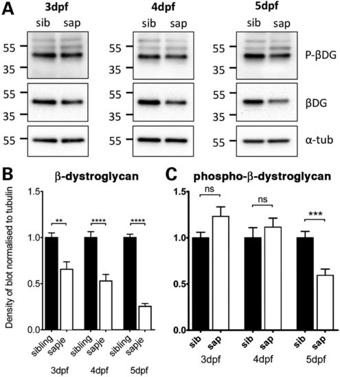
Levels of β-dystroglycan and β-dystroglycan phosphorylated on tyrosine in sapje and sibling larvae. Western blots of lysates of individual 3, 4 and 5 dpf sibling and sapje larvae western blotted with antibodies against phosphorylated β-dystroglycan [p-β-DG, (A) top], non-phosphorylated β-dystroglycan [β-DG, (A) middle] and α-tubulin was used as a loading control [α-tub, (A) bottom]. Numbers represent relative position of molecular weight markers in kDa. (B and C) The integrated density of the blots probed against β-DG and p-β-DG shown in (A), quantified relative to α-tubulin levels in each sample. Graphs show mean + SEM of 12 (B) or 9 (C) samples from three independent experiments in each case. (B) There is a significant decrease in the level of β-dystroglycan in larvae with the sapje mutation at 3, 4 and 5 dpf (unpaired t-tests, 3 dpf: P = 0.0016; 4 dpf: P<0.0001; 5 dpf P < 0.0001). (C) Levels of phosphorylated dystroglycan are slightly increased in sapje at 3 and 4 dpf, but this increase is not statistically significant (P > 0.05). When compared with sibling lysates, levels of phosphorylated dystroglycan at 5 dpf are significantly lower in sapje (P = 0.0007). EXPRESSION / LABELING:
PHENOTYPE:
|
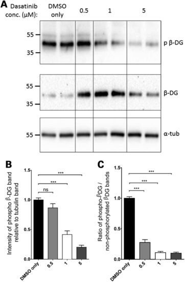
Phosphorylated β-dystroglycan in dasatinib-treated wild-type zebrafish larvae. Pools of 10 LWT zebrafish embryos were treated with dasatinib or DMSO from 24 hpf until 96 hpf and then lysed for SDS–PAGE. (A) A western blot of lysates probed with antibodies against phosphorylated β-dystroglycan (top panel), β-dystroglycan (middle panel) and α-tubulin (bottom panel). Numbers represent relative position of molecular weight markers in kDa. (B) The density of the blot probed against phosphorylated dystroglycan was quantified relative to α-tubulin levels in each sample, and represented as a ratio to the average DMSO only control signal. There is a significant decrease in the level of phosphorylated β-dystroglycan in dasatinib-treated embryos, compared with DMSO-treated controls. (C) The density of the blot probed against phosphorylated dystroglycan was quantified relative to non-phosphorylated dystroglycan levels in each sample, and normalized to the average DMSO only control signal. There is a significant decrease in the ratio of phosphorylated to non-phosphorylated dystroglycan in dasatinib-treated embryos, compared with DMSO-treated controls. Graphs show mean + SEM of at least eight samples for each treatment, from three independent experiments. One-way analysis of variance (ANOVA) was carried out followed by Dunnett′s multiple comparison test (ns, non-significant; ***P < 0.001). |

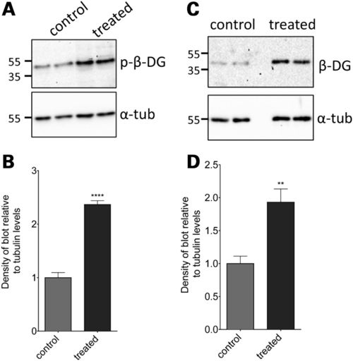
The effect of dasatinib treatment on levels of phosphorylated and non-phosphorylated β-dystroglycan in sapje zebrafish larvae. Lysates were made from sapje–/– embryos treated from 24 hpf until 96 hpf with dasatinib or DMSO-only. (A) Western blots probed with antibodies for p-β-DG and α-tubulin. (B) The density of the blot probed for p-β-DG was quantified relative to α-tubulin levels in each sample, and normalized to the average control signal. There is a significant decrease in the level of phosphorylated β-dystroglycan in larvae treated with dasatinib, compared with controls. (C) Western blots probed with antibodies for β-DG and α-tubulin. Numbers in (A and C) represent relative position of molecular weight markers in kDa. (D) The density of the blot probed against β-DG was quantified relative to α-tubulin levels in each sample, and normalized to the average control signal. There was a significant difference in the levels of β-DG in larvae between different treatment groups. Graphs show mean + SEM of at least six samples for each treatment, from three independent experiments. One-way ANOVA was carried out followed by Dunnett′s multiple comparison test (ns, non-significant; ***P < 0.001). |
The effect of MG132 treatment on levels of phosphorylated and non-phosphorylated β-dystroglycan in sapje zebrafish larvae. Lysates were made from sapje/ embryos treated from 24 hpf until 96 hpf with 5 μM MG132 or DMSO-only. (A) Western blots probed with antibodies for β-dystroglycan (β-DG) and α-tubulin (α-tub). (B) The density of the blot probed with β-DG was quantified relative to α-tubulin levels in each sample, and normalized to the average control signal. There was a significant increase in the level of β-dystroglycan in larvae treated with MG132, compared with controls (unpaired t-test: t = 4.048, df = 10, P = 0.0023). (C) Western blots probed with antibodies for phosphorylated β-dystroglycan (p-β-DG) and α-tubulin. (D) The density of the blot probed with p-β-DG was quantified relative to α-tubulin levels in each sample, and normalized to the average control signal. There was a significant increase in the levels of β-DG in larvae between different treatment groups (unpaired t-test: t = 11.47, df = 10, P < 0.0001). Graphs represent the mean of six samples from three independent experiments, error bars are SEM. |
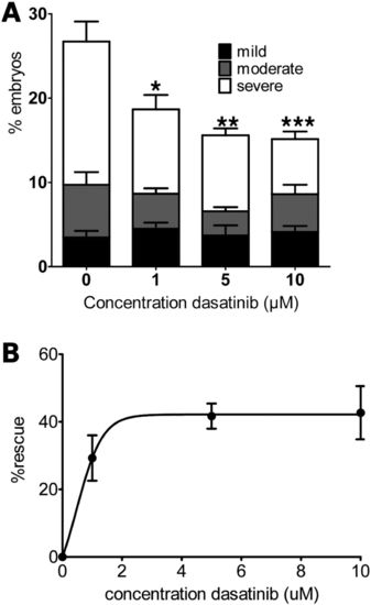
The effect of 72 h dasatinib treatment on sapje muscle phenotype. Embryos were treated with dasatinib or DMSO only for 72 h before a birefringence assay was carried out. The number of larvae showing normal or disrupted muscle birefringence was counted. (A) The proportion of larvae showing mild, moderate and severe muscle damage. There was a significant decrease in the percentage of larvae showing a severe phenotype in the groups treated with dasatinib compared with DMSO alone. (B) The overall percentage rescue of the dystrophic phenotype is plotted against dasatinib concentration. Data are plotted taking the proportion of dystrophic fish in DMSO-only treated groups as 0% rescue (one-way ANOVA followed by Dunnett′s multiple comparison test: *P < 0.05, **P < 0.01 and ***P < 0.001). Data points represent mean values of five independent experiments, with a total of <250 embryos per treatment group. Error bars represent SEM. PHENOTYPE:
|
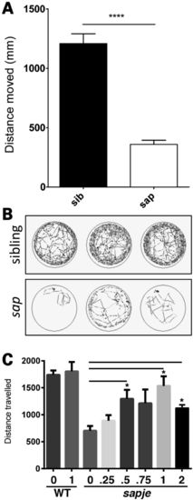
Dasatinib treatment rescues swimming ability in sapje larvae. Using ViewPoint video tracking and analysis, the overall distance moved by sapje larvae is significantly lower compared with wild-type siblings (A, P < 0.0001). Each bar represents mean tracking data from 36 larvae from three separate multiwell plates, and error bars represent SEM. (B) Representative traces for three sapje (sap, bottom) and three sibling larvae (top), light grey traces represent periods of fast movement and dark grey periods of slow movement. (C) Sapje and control larvae were treated with the indicated concentrations of dasatinib (μM), or DMSO only, from 3 to 5 dpf. Dasatinib has a dose-dependent effect in increasing swimming duration compared with vehicle-alone up to a maximum concentration of 1 μM. One micromolar dasatinib had no effect on swimming activity of wild-type zebrafish. One-way ANOVA analysis of ViewPoint tracking data indicated a significant increase between dasatinib-treated and control groups for the distance moved (one-way ANOVA: F = 3.188, df = 2.69, P = 0.0474, followed by Dunnett′s multiple comparison test: *P < 0.05, ns = non-significant). PHENOTYPE:
|