
- Title
-
PEG-PLA Nanoparticles facilitate siRNA knockdown in adult zebrafish heart
- Authors
- Diao, J., Wang, H., Chang, N., Zhou, X.H., Zhu, X., Wang, J., Xiong, J.W.
- Source
- Full text @ Dev. Biol.
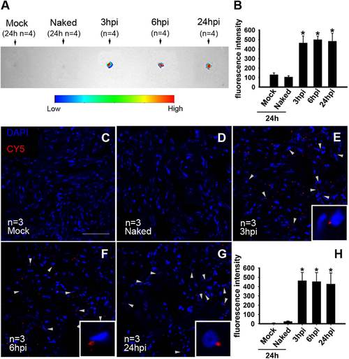
Effective nanoparticle-assisted delivery of siRNA into the adult zebrafish heart. (A) Fluorescence images of CY5-siRNA accumulation in isolated zebrafish heart under the in vivo imaging system. Note that fluorescence signals were hardly detectable in the negative control (Mock) and unencapsulated siRNA (Naked) groups at 24 hpi, and were increased in the nanoparticle-encapsulated siRNA groups from 3 to 24 hpi. An arbitrary scale of fluorescence signal is shown from blue (low) to red (high). (B) Quantification of CY5-siRNA in heart tissues using the in vivo imaging system (*P<0.05). (C–G) Fluorescent images of cryosections of heart tissues under confocal microscopy. Note the clear CY5-siRNA signal in the experimental group at 3, 6 and 24 hpi but very little signal in the negative control (Mock) and naked siRNAs (Naked) groups at 24 hpi. Enlarged siRNA localization is shown (E–G, low-right corner). DAPI was used to stain nuclei. Scale bar, 50 µm. (H) Quantification of the CY5-siRNA fluorescence signals in Fig. 2C–G. (*P<0.05). |

Efficient siRNA knockdown of aldh1a2 and dusp6 in the adult zebrafish heart. (A, B) Immunostaining showed Aldh1a2 expression in the epicardium and endocardium of negative control (siN.C.)-injected hearts (A, A′) and siAldh1a2 siRNA-injected hearts (B, B′). MF20 staining marks the myocardium. (C, D) Western blots showing that nanoparticle-encapsulated siAldh1a2 reduced Aldh1a2 expression to a level comparable to that in the sham group, compared to the nanoparticle-encapsulated siN.C. and naked siAldh1a2 groups at 2 dpa (C), and quantitative knockdown of Aldh1a2 (D). (E, F) Immunostaining showed Dusp6 expression in the epicardium of siN.C.-injected control hearts (E, E′) and siDusp6-injected hearts (F, F′). Mef2C staining marks the myocardium. (G, H) Western blots showing that nanoparticle-encapsulated siDusp6 reduced Dusp6 expression to a level comparable to that in the sham group, compared with the nanoparticle-encapsulated siN.C. and naked siDusp6 groups at 2 dpa (G), and quantitative knockdown of Dusp6 (H). Scale bars, 100 µm. |

Nanoparticle-aided suppression of Aldh1a2 impedes cardiac regeneration. (A, B) siAldh1a2 inhibited Gata4-GFP cardiomyocytes in the injured areas (B, B′) compared with negative (siN.C.) control treatment (A, A′). MF20 marks the myocardium. (C, D) siAldh1a2 diminished the numbers of BrdU+/Mef2C+ proliferating myocytes (D, D′) compared with siN.C. treatment (C, C′) at 14 dpa. (E) Statistics of Gata4-GFP cardiomyocytes from A, B. (F) Statistics of Mef2C+/BrdU+ proliferating myocytes from C, D. (C′, C′′, C′′′) High-magnification images of the squared image in panel C showing Mef2C+/BrdU+ colocalization (C′), red fluorescent channel for Mef2C (C′′) and green fluorescent channel for BrdU (C′′′). (D′, D′′, D′′′) High-magnification images of the squared image in panel D showing Mef2C+/BrdU+ colocalization (D′), red fluorescent channel for Mef2C (D′′) and green fluorescent channel for BrdU (D′′′). *P<0.05; scale bars for A–D, 100 µm. |
Reprinted from Developmental Biology, 406(2), Diao, J., Wang, H., Chang, N., Zhou, X.H., Zhu, X., Wang, J., Xiong, J.W., PEG-PLA Nanoparticles facilitate siRNA knockdown in adult zebrafish heart, 196-202, Copyright (2015) with permission from Elsevier. Full text @ Dev. Biol.