
- Title
-
Invasiveness and metastasis of retinoblastoma in an orthotopic zebrafish tumor model
- Authors
- Chen, X., Wang, J., Cao, Z., Hosaka, K., Jensen, L., Yang, H., Sun, Y., Zhuang, R., Liu, Y., Cao, Y.
- Source
- Full text @ Sci. Rep.
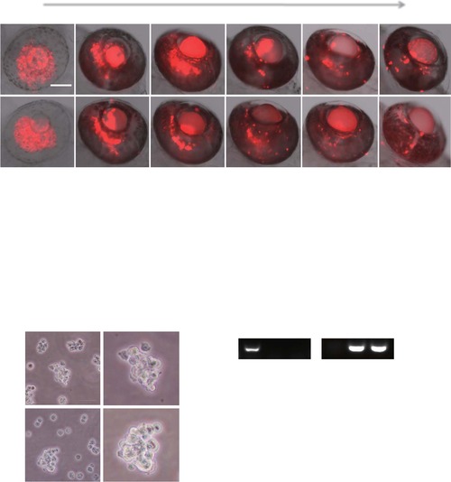
Formation of primary retinoblastoma in the zebrafish retina, and re-culture of implanted retinoblastoma cells from zebrafish in vitro. (a) Approximately, 150 DiI-labeled human (RB355) or mouse (SJmRBL-8) retinoblastoma cells (red color) were intravitreally implanted into each eye of zebrafish and the formation of primary tumors were kinetically monitored under fluorescent microscopy at different time points after tumor cell implantation. Bar = 100 µm. (b) Primary tumor areas were quantified using a digital Photoshop program. Approximately 30–40 tumor samples were used for quantification. (c) Transplanted SJmRBL-8 retinoblastoma cells are successfully isolated and cultured in vitro. The photographs at day 14 from inverted microscope showed the transplanted cells maintained their morphology and viability after injection. (d) Total RNA was extracted from zebrafish embryos, mouse SJmRBL-8 cells and the re-cultured cells, and RT-PCR was then used to analyze zebrafish and mouse GAPDH expression. (e) The expression of zebrafish and mouse GAPDH was determined by quantitative real-time PCR. |

Retinoblastoma invasion and metastasis in the zebrafish (a) Time-course monitoring invasion and metastasis of DiI-labeled mouse SJmRBL-8 retinoblastoma cells (red color) in the head region of zebrafish embryos. Arrows indicate metastatic tumor cells in single cell or a cluster cells. Bar = 100 µm. (b) Quantification of percentage of zebrafish embryos with contralateral ocular SJmRBL-8 metastasis, percentage of zebrafish embryos with head metastasis, percentage of zebrafish embryos with trunk metastasis, and the average total number of metastatic tumor cells per zebrafish (n = 40 embryos/group). (c) Time-course monitoring invasion and metastasis of DiI-labeled human RB355 retinoblastoma cells (red color) in the head region of zebrafish embryos. Arrows indicate metastatic tumor cells in single cell or a cluster cells. Bar = 100 µm. (d) Quantification of percentage of zebrafish embryos with contralateral ocular RB355 metastasis, percentage of zebrafish embryos with head metastasis, percentage of zebrafish embryos with trunk metastasis, the average total number of metastatic tumor cells per zebrafish, and the distribution of metastatic SJmRBL-8 and RB355 tumor cells in head and trunk regions at day 4 after primary tumor implantation (n = 40 embryos/group). |

Interaction between retinoblastoma cells and vasculatures (a) A representative image of the entire fli1:EGFP zebrafish embryo that received SJmRBL-8 retinoblastoma cell implantation at day 4. The left eye was implanted with DiI-labeled tumor cells (red) and zebrafish vasculatures were visualized by expression of EGFP. Arrows point to the metastatic tumor cells in the head and trunk regions. Bar = 100 µm. (b) Detection of DiI-labeled metastatic retinoblastoma cells in the contralateral eye by confocal imaging analysis. (c) Visualization of implanted retinoblastoma cells in relation to hyaloid vasculatures at different points after tumor cell implantation. Arrows indicate the position of primary tumors growing in tight association with hyaloid vessels. Bar = 100 µm. |

Vegf-aa morpholino treatment attenuates retinoblastoma metastasis. (a) Vegf-aa specific morpholinos and control morpholinos were injected into the yolk of each embryo at the 1-2 cell stage. DiI-labeled SJmRBL-8 retinoblastoma cells were intravitreally implanted in zebrafish at 48 hpf. Retinoblastoma invasion and metastasis were monitored at day 0 and day 4 post-implantation. Arrows point to metastatic tumor cells. Bar = 100 µm. (b) Quantification of metastatic tumor cells and the averages of maximal distance of metastatic foci in Vegf-aa and control morpholinos-treated zebrafish embryos (n = 20 embryos/group). |

Sunitinib inhibits mouse retinoblastoma invasion and metastasis. (a) SJmRBL-8 retinoblastoma cells were intravitreally implanted in zebrafish and sunitinib was added to the aquarium water to constitute a final concentration of 1 µM. Retinoblastoma invasion and metastasis were monitored at different time points. Arrows point to metastatic tumor cells. Bar = 100 µm. (b) Quantification of metastatic tumor cells and the averages of maximal distance of metastatic foci in vehicle- and sunitinib-treated zebrafish embryos (n = 60 embryos/group). |
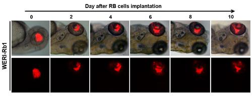
Formation of primary retinoblastoma, invasion and metastasis of non-invasive retinoblastoma cells in the zebrafish. Approximately 150 DiI-labeled human non-invasive WERI-Rb1 retinoblastoma cells (red color) were intravitreally implanted into the eye of zebrafish embryo. The formation of primary tumors, and invasion and metastasis were kinetically monitored under fluorescent microscopy at different time points after tumor cell implantation. Approximately 30-40 zebrafish were used for this experiment. |

Sunitinib inhibits human retinoblastoma invasion and metastasis. (a) RB355 retinoblastoma cells were intravitreally implanted in zebrafish and sunitinib was added to the aquarium water to constitute a final concentration of 1 . Retinoblastoma invasion and metastasis were monitored at different time points. Arrows point to metastatic tumor cells. Bar = 100 µm (b) Quantification of metastatic tumor cells and the averages of maximal distance of metastatic foci in vehicle- and sunitinib-treated zebrafish embryos (n = 60 embryos/group). |