
- Title
-
Two types of transgenic lines for doxycycline-inducible, cell-specific gene expression in zebrafish ultraviolet cone photoreceptors
- Authors
- West, M.C., Campbell, L.J., Willoughby, J.J., and Jensen, A.M.
- Source
- Full text @ Gene Expr. Patterns
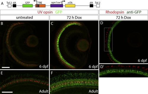
Generation of an UV cone-specific, doxycycline-inducible, self-reporting gene expression system. (A) Diagram of the construct used to generate the stable, transgenic line Tg(opn1sw1:rtTA, TRE:GFP), which expresses doxycycline (Dox)-inducible green fluorescent protein (GFP) specifically in UV cone photoreceptors. The UV opsin promoter (opn1sw1) drives expression of the reverse tetracycline-controlled transcriptional activator (rtTA) gene while the tetracycline responsive element (TRE) is upstream of GFP. (B, C) Confocal z-projections of retinal sections from 6 dpf Tg(opn1sw1:rtTA, TRE:GFP); alb/ larvae labeled with an anti-UV opsin antibody (red). (B) GFP fluorescence (green) is not visible in the UV cones of larval transgenic zebrafish in the absence of Dox treatment. (C) GFP fluorescence is clearly visible in UV cones in transgenic larvae after 72 h of Dox treatment (3–6 dpf). (D) Confocal z-projection of retinal section from 6 dpf Tg(opn1sw1:rtTA, TRE:GFP); alb/ larva labeled with an anti-GFP antibody (green) and an anti-Rhodopsin antibody (red). (D2) Corresponds to boxed area in D. Anti-GFP immunofluorescence (green) is visible in UV opsin cone photoreceptors while anti-Rhodopsin immunofluorescence (red) is visible in the outer segments of rod photoreceptors. (E, F) Confocal z-projections of the photoreceptor layer of retinas from adult Tg(opn1sw1:rtTA, TRE:GFP) zebrafish labeled with anti-UV opsin antibody (red). (E) GFP fluorescence (green) is not visible in the adult transgenic UV cones in the absence of Dox treatment. (F) GFP fluorescence is clearly visible in transgenic adult UV cones after 72 h of Dox treatment. dA, polyadenylation signal; Tol2, pTol integration site. Scale bars (B, E), 50 μm. |

Time course of doxycycline-induced GFP expression in Tg(opn1sw1:rtTA, TRE:GFP); alb/ larvae. Confocal z-projections of retinal sections from 6 dpf Tg(opn1sw1:rtTA, TRE:GFP); alb/ larvae that were (A) untreated or treated with 10 μg/mL Dox for (B) 2, (C) 4, (D) 8, (E) 16, (F) 24, (G) 48, or (H) 72 h. (A–H) Anti-UV opsin immunofluorescence (red) labels UV cone outer segments. GFP fluorescence (green) is apparent after 8 h of treatment (D, D inset) and continues to increase by number of cones with the increase in Dox exposure time (E–H). (I) Anti-GFP immunofluorescence (green) in untreated transgenic larvae indicates GFP expression that is not present in untreated, wild type larvae (J). Scale bar (A), 50 μm. |
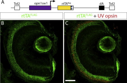
Generation of UV-cone-specific, epitope-tagged, rtTA-expressing transgenic line for doxycycline-inducible gene expression. (A) Diagram of the construct used to generate the stable transgenic zebrafish line, Tg(opn1sw1:rtTAflag); alb/, which expresses a FLAG-tagged rtTA protein specifically in UV cone photoreceptors. The UV opsin promoter (opn1sw1) drives expression of the flag-epitope tagged reverse tetracycline-controlled transcriptional activator (rtTAflag) gene. (B, C) Confocal z-projection of a retinal section from a 6 dpf Tg(opn1sw1:rtTAflag); alb/ larva labeled with anti-FLAG and anti-UV opsin antibodies. (B) Anti-FLAG immunofluorescence (green) is visible in the photoreceptor layer of the retina. (C) Anti-UV opsin immunofluorescence (red) colocalizes with the anti-FLAG immunofluorescence (green) in UV opsin cone photoreceptors. dA, polyadenylation signal; Tol2, pTol integration site. Scale bar (C), 50 μm. |
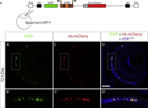
Bidirectional transactivation of an injected biTRE-containing plasmid into Tg(opn1sw1:rtTAflag). (A) Diagram of the bidirectional tetracycline response element (biTRE)-containing construct injected into Tg(opn1sw1:rtTAflag) one-cell embryos. EGFP and mCherry with a nuclear localization sequence (nls-mCherry) flank the biTRE. (B–D) Confocal z-projection of a retinal section from an injected 6 dpf Tg(opn1sw1:rtTAflag) larva treated with Dox for 72 h and labeled with anti-FLAG antibody (blue). GFP fluorescence (B, B2 green) and nls-mCherry fluorescence (C, C2 red) are visible in the photoreceptor layer and co-localize with anti-FLAG immunofluorescence (D, D2 blue) in UV cones. Boxed regions in B, C, and D correspond to B2, C2, and D2, respectively. dA, polyadenylation sequence; Tol2, pTol integration site. Scale bar (D), 50 μm. |
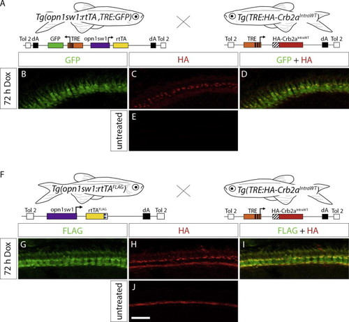
Transactivation in the Tg(TRE:HA-Crb2aIntraWT) line. (A) Diagram indicating the cross of Tg(opn1sw1:rtTA, TRE:GFP) to Tg(TRE:HA-Crb2aIntraWT) for double-transgenic embryo production. (B–D) Confocal z-projection of a retinal section from a 6 dpf Tg(opn1sw1:rtTA, TRE:GFP; TRE:HA-Crb2aIntraWT) larva treated with Dox for 72 h and labeled with anti-HA antibody. (B) GFP fluorescence (green) and (C) anti-HA immunofluorescence (red) indicate that HA-Crb2aIntraWT is expressed in UV cones (D, merge, yellow). (E) Confocal z-projection of an untreated 6 dpf Tg(opn1sw1:rtTA, TRE:GFP; TRE:HA-Crb2aIntraWT) retinal section labeled with anti-HA antibody. (F) Diagram indicating the cross of Tg(opn1sw1:rtTAflag) to Tg(TRE:HA-Crb2aIntraWT) for double-transgenic embryo production. (G–I) Confocal z-projection of a retinal section from a 6 dpf Tg(opn1sw1:rtTAflag; TRE:HA-Crb2aIntraWT) larva treated with Dox for 72 h and labeled with anti-FLAG and anti-HA antibodies. (G) Anti-FLAG immunofluorescence (green) and (H) anti-HA immunofluorescence (red) indicate HA-Crb2aIntraWT is expressed in the UV cones (I, merge, yellow). (J) Confocal z-projection of an untreated retinal section from a 6 dpf Tg(opn1sw1:rtTAflag; TRE:HA-Crb2aIntraWT) larva labeled with anti-HA antibody. Scale bar (J), 10 μm. |
Reprinted from Gene expression patterns : GEP, 14(2), West, M.C., Campbell, L.J., Willoughby, J.J., and Jensen, A.M., Two types of transgenic lines for doxycycline-inducible, cell-specific gene expression in zebrafish ultraviolet cone photoreceptors, 96-104, Copyright (2014) with permission from Elsevier. Full text @ Gene Expr. Patterns