
|
Fig. 5
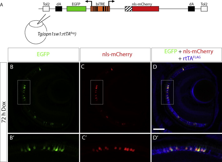
Bidirectional transactivation of an injected biTRE-containing plasmid into Tg(opn1sw1:rtTAflag). (A) Diagram of the bidirectional tetracycline response element (biTRE)-containing construct injected into Tg(opn1sw1:rtTAflag) one-cell embryos. EGFP and mCherry with a nuclear localization sequence (nls-mCherry) flank the biTRE. (B–D) Confocal z-projection of a retinal section from an injected 6 dpf Tg(opn1sw1:rtTAflag) larva treated with Dox for 72 h and labeled with anti-FLAG antibody (blue). GFP fluorescence (B, B2 green) and nls-mCherry fluorescence (C, C2 red) are visible in the photoreceptor layer and co-localize with anti-FLAG immunofluorescence (D, D2 blue) in UV cones. Boxed regions in B, C, and D correspond to B2, C2, and D2, respectively. dA, polyadenylation sequence; Tol2, pTol integration site. Scale bar (D), 50 μm.
Reprinted from Gene expression patterns : GEP, 14(2), West, M.C., Campbell, L.J., Willoughby, J.J., and Jensen, A.M., Two types of transgenic lines for doxycycline-inducible, cell-specific gene expression in zebrafish ultraviolet cone photoreceptors, 96-104, Copyright (2014) with permission from Elsevier. Full text @ Gene Expr. Patterns