
- Title
-
A synthetic dl-nordihydroguaiaretic acid (Nordy), inhibits angiogenesis, invasion and proliferation of glioma stem cells within a zebrafish xenotransplantation model
- Authors
- Yang, X., Cui, W., Yu, S., Xu, C., Chen, G., Gu, A., Li, T., Cui, Y., Zhang, X., and Bian, X.
- Source
- Full text @ PLoS One
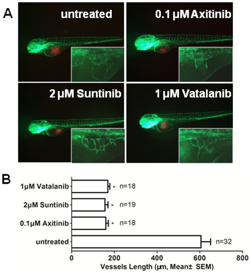
The effects of verified VEGF receptor tyrosine kinase inhibitor on angiogenesis that was induced by GSCs within zebrafish embryos. A. The representative images of the inhibition of angiogenesis with axitinib, suntinib and vatalanib treatment. B. Quantitative analysis of the length of newly formed vessels that were induced by U87 GSCs with/without axitinib, suntinib and vatalanib treatment at 2 dpi. (P<0.0001) PHENOTYPE:
|
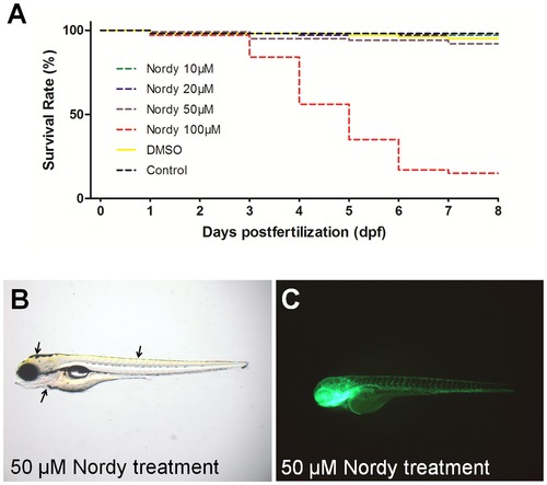
The response to Nordy treatment on the survival rate, and development of zebrafish embryos. A. Quantitative analysis of the survival rate of naïve zebrafish embryos following treatment with various concentrations of Nordy. B. Showing the representative bright field images of native zebrafish embryos that were incubated with 50 μM Nordy. C. The representative images of vascular development of Tg (fli1:EGFP)y1 embryos following treatment with 50 μM Nordy. |
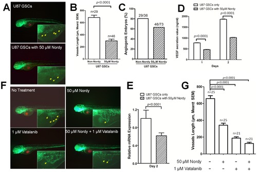
Analysis of angiogenesis induced by U87 GSCs with/without Nordy treatment. A. The representative merged images of angiogenesis as induced by U87 GSCs with/without Nordy treatment. The images at higher magnification show the new vessels that were induced by U87 GSCs. B. Quantitative analysis of the length of newly formed vessels that were induced by U87 GSCs with/without Nordy treatment at 2 dpi. C. Quantitative analysis of the percentage of angiogenic embryos with/without Nordy treatment at 2 dpi. D. Showing the secreted VEGF165 volume of U87 GSCs with/without Nordy treatment in vitro. E. Showing the VEGF165 mRNA level in U87 GSCs with/without Nordy treatment as assayed by qRT-PCR at 2 dpi. F. Showing the representative merged images of angiogenesis that were induced by U87 GSCs treated with/without Nordy and/or vatalanib. The images at higher magnification showed new vessels induced by tumor cells. G. Showing quantitative analysis of the length of newly formed vessels induced by U87 GSCs with/without Nordy and/or Vatalanib treatment. The yellow arrow indicates newly formed angiogenic vessels on embryonic yolk sac ball. Red: injected RFP-labeled U87 GSCs cells; green: GFP fluorescence of vasculature in Tg (fli1:EGFP)y1 embryos. |

CD133 positive U87 GSCs were suppressed by Nordy treatment within zebrafish embryos. A. The representative high-invasion phenotypes of injected GSCs with/without Nordy treatment. B. The inhibitory effect of Nordy on the invasion of CD133 positive U87 GSCs within zebrafish embryos. The percentages of invasive cells in adoptively transferred embryos (low, medium, or high-invasion) were measured at 2 dpi. The data were obtained from three replicate experiments with the number of embryos: n = 120 for the non-Nordy treated group, n = 113 for the Nordy treated group. C. The relationship between angiogenesis induced by GSCs and degree of invasion. N = 34 represented the low-invasion group, n = 33 represented the medium-invasion group, and n = 39 represented the high-invasion group. Red: adoptively transferred RFP-labeled U87 GSCs cells; green: represents the GFP fluorescence of angiogenesis in Tg (fli1:EGFP)y1 embryos. PHENOTYPE:
|

Effects of Nordy on the proliferation of GSCs within zebrafish embryos. A. Representative merged images of GSC proliferation with/without Nordy treatment within zebrafish embryos. B. Quantitative analysis of the emitted fluorescence of RFP-labeled GSCs with/without Nordy treatment at 2 dpi and 4 dpi. At higher magnification, the images showed the injected the RFP labeled GSCs accumulate within the embryos (yellow broken box). Red: injected U87-RFP cells; green: GFP fluorescence of angiogenesis in Tg (fli1:EGFP)y1 embryos. |
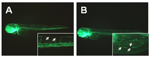
Vascular disruption phenotypes. A. Showing intersegmental blood vessel disruption. B. Showing subintestinal vein developmental disruption. |
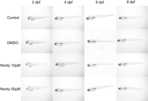
The morphology of zebrafish embryos incubated with 10 μM, and 50 μM Nodry as compared to the morphology of normal embryos. |

Angiogenesis induced by differentiated U87 cells and U87 in GSC in zebrafish. A: Representative merged images of angiogenesis induced by differentiated U87 cells and U87 GSCs in zebrafish embryos. The images here are at a higher magnification and showed new vessels that were induced by tumor cells. B. Quantitative analysis of the length of newly formed vessels induced by differentiated U87 cells and U87 GSCs in zebrafish embryos. C. Quantitative analysis of the percentage of angiogenic embryos induced by differentiated U87 cells and U87 GSCs. |

Time-lapse merged images of invasive U87 GSCs with/without Nordy treatment within zebrafish embryos (6 hpi, 18 hpi, 30 hpi, 42 hpi, and 54 hpi). |