
- Title
-
Fermitins, the Orthologs of Mammalian Kindlins, Regulate the Development of a Functional Cardiac Syncytium in Drosophila melanogaster
- Authors
- Catterson, J.H., Heck, M.M., and Hartley, P.S.
- Source
- Full text @ PLoS One
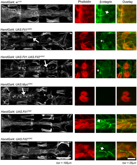
The monochrome micrographs show the adult Drosophila heart (segments A2 to A5) stained with phalloidin, oriented with the anterior region to the left. Genes were silenced using the Hand-Gal4 enhancer which drives expression in cardiomyocytes (as well as pericardial nephrocytes and enterocytes of the gut) and abnormal cardiomyocyte/heart morphology is highlighted by the arrows. The colour panels show higher magnification micrographs of at least two cardiomyocytes stained with phalloidin (red) and antibodies to the Drosophila β-integrin, myospheroid (green); arrowheads indicate normal integrin staining between cardiomyocytes; asterisks indicate an abnormal staining pattern associated with loss of cardiomyocyte junction integrity. A wild type phenotype (Hand-Gal4; w1118), is characterised by contiguous cardiomyocytes and β-integrin staining between cardiomyocytes. |
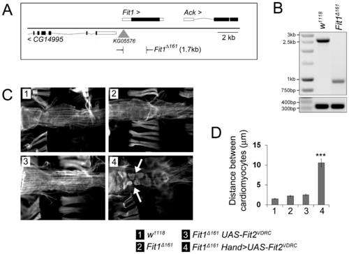
Cardiomyocyte Fit2 compensates for the loss of Fit1 to establish the cardiac syncytium. (A) Schematic showing the Fit1 locus with the adjacent genes (CG14995 and Ack) and the site of the P-element insertion used to create the 1.7 kb deletion mutant (Fit1Δ161). (B) PCR of genomic DNA from wild-type (w1118) and Fit1Δ161 mutant flies. (C) Micrographs of adult hearts stained with phalloidin. The arrow highlights an abnormal cardiomyocyte phenotype. (D) Mean (±SEM) distance between neighbouring cardiomyocytes. n = 12–16 measurements from four to seven independent flies for each genotype. ***P<0.001 from all other genotypes. Legend applies to C & D. |