
- Title
-
A spatio-temporal model of notch signalling in the zebrafish segmentation clock: conditions for synchronised oscillatory dynamics
- Authors
- Terry, A.J., Sturrock, M., Dale, J.K., Maroto, M., and Chaplain, M.A.
- Source
- Full text @ PLoS One
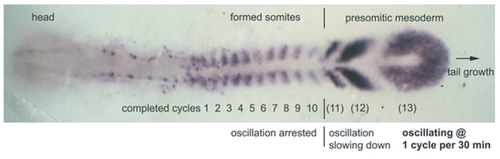
Top view micrograph of formed and forming somites in a zebrafish embryo. The embryo is at 10-somite stage, stained by in situ hybridization for deltaC mRNA. The deltaC gene exhibits oscillatory expression in the presomitic mesoderm (PSM). The oscillations are quickest in cells at the tail end (posterior) PSM. Cells in the posterior PSM enter the anterior PSM and then the somitic mesoderm (the region of formed somites) as the tail end of the embryo grows away from them. As the tail end grows it releases a Wnt and FGF signal. This gives rise to a morphogen gradient, which causes the oscillations to slow down in cells the further they are from the tail end. Cells cease oscillating altogether and form somites as they exit the anterior PSM. The spatio-temporal expression pattern of the her1 and her7 genes is very similar to the expression pattern for deltaC. Reproduced from figure 1a in [5] under the Creative Commons Attribution License. EXPRESSION / LABELING:
|