
- Title
-
Conservation, expression, and knockdown of zebrafish plxnb2a and plxnb2b
- Authors
- Perälä, N., Peitsaro, N., Sundvik, M., Koivula, H., Sainio, K., Sariola, H., Panula, P., and Immonen, T.
- Source
- Full text @ Dev. Dyn.

Expression of plxnb2a in 24 hours postfertilization (hpf) to 4 days postfertilization (dpf) zebrafish embryos by whole-mount in situ hybridization. A-D: Lateral views. E-H: Dorsal views. I: Lateral view of a 3 dpf embryo. J-V′: Transverse sections of 2 dpf (J-O), 3 dpf (P-U), and 24 hpf (V,V′) whole-mount samples. X: No staining was seen with the control sense probe. Y,Z: Sagittal sections of 2 dpf (Y) and 3 dpf (Z) whole-mount samples. Ce, cerebellum; Cm, cranial mesenchyme; Eth, ethmoid plate; Fg, facial ganglion; Io, neuromasts of the infraorbital lateral line; Ll, lateral line ganglia; Mhb, midbrain-hindbrain boundary; Mo, medulla oblongata; N, notochord; Ov, otic vesicle; Pa, pharyngeal arches; Pd, pronephric duct; Pf, pectoral fin; Pha, pharynx; Pq, palatoquadrate; RhV, rhombencephalic ventricle; S, somite; So, neuromasts of the supraorbital lateral line. Scale bars = 100 μm in J-V′,Y-Z. |

Expression of plxnb2b in 24 hours postfertilization (hpf) to 4 days postfertilization (dpf) zebrafish embryos by whole-mount in situ hybridization. A-D: Lateral views. E-H: Dorsal views. I: Lateral view of a 3 dpf embryo. J-U: Transverse sections of J-O at 2 dpf and P-U at 3 dpf whole-mount samples. V,X: Sagittal sections of 2 dpf (V) and 3 dpf (X) whole-mount samples. Y: No staining was seen with the control sense probe. Allg, anterior lateral line ganglion; Ce, cerebellum; Cm, cranial mesenchyme; Eth, ethmoid plate; Fg, facial ganglion; Mhb, midbrain-hindbrain boundary; Mo, medulla oblongata; N, notochord; Ob, olfactory bulb; Ov, otic vesicle; P, pallium; Pa, pharyngeal arches; Pd, pronephric duct; Pf, pectoral fin; Pha, pharynx; RhV, rhombencephalic ventricle; S, somite; So, neuromasts of the supraorbital lateral line; TeV, telencephalic ventricle; Th, thalamus; Vg, vagal ganglion. Scale bars = 100 μm in J-X. |
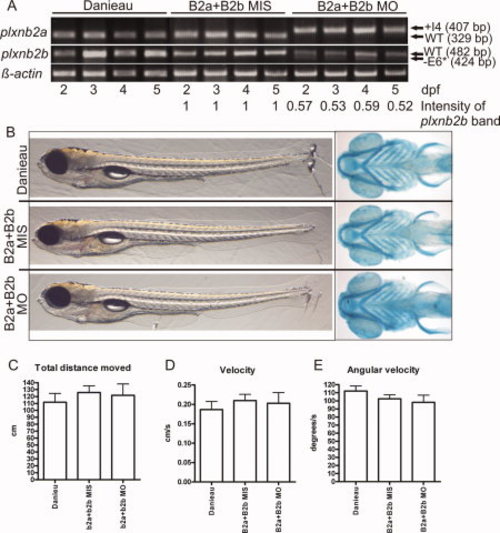
Normal development and locomotion of double plxnb2a and plxnb2b morphants. A: Reverse transcriptase-polymerase chain reaction (RT-PCR) analysis of the efficiency of the double knockdown using B2aMO and B2bMO morpholinos interfering with the splicing. Each morpholino was used at a concentration of 5 ng per embryo. E6* = 32 end of exon 6. B: Light microscopy images of lateral views of the double morphant as well as the controls at 5 days postfertilization (dpf). Ventral views of Alcian blue-stained 5 dpf double morphant and controls visualizing the cartilage structure. C-E: Results of the swimming performance assay from one representative replicate (zebrafish larvae per group n = 16-27). Graphs of the total distance moved (C), velocity (D), and angular velocity (E) are represented as mean and standard error of mean. |
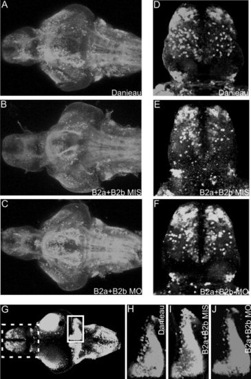
The GABAergic neurons in 5 dpf zebrafish brain. A-C: A horizontal overview of stacked confocal scans from the ventral side of the brain after treatment with Danieau (A), B2aMIS+B2bMIS (5 ng+5 ng; B), or B2aMO+B2BMO (5 ng+5 ng; C). D-F: Dorsal view of the telencephalon after the injection of Danieau (D), B2aMIS+B2bMIS (E), or B2aMO+B2bMO (F) treatment, showing the GABAergic neurons in the olfactory bulb and medial telencephalon. G: Dorsal overview of the GABAergic neurons in the 5-day zebrafish brain. The dashed box (telencephalon) corresponds to the area enlarged in D-F. The cerebellar GABAergic neurons are boxed. H-J: Enlargements of the cerebellar GABAergic neuron area (boxed in G) of Danieau- (H), B2aMIS+B2bMIS- (I), and B2aMO+B2bMO- (J) treated samples. |
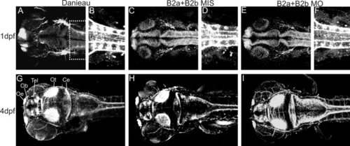
Whole-mount staining with anti-acetylated α-tubulin antibody to view axon projections in 1 days postfertilization (dpf) and 4 dpf zebrafish. A-F: Confocal scans of 1dpf zebrafish embryos. A: A dorsal view of the head of a Danieau-treated embryo. B: A ventral view of the spinal axons (area boxed in A). C-F: B2aMIS+B2bMIS-treated (5 ng+5 ng, C,D) and B2aMO+B2BMO-treated (5 ng+5 ng, E,F) embryos. G–I: Ventral stacks of 4 dpf zebrafish embryo head after Danieau-treatment (G), B2a+B2bMIS treatment (H), and B2aMO+B2bMO treatment (I). Ce, cerebellum; Ob, olfactory bulb; Oe, olfactory epithelium; Ot, optic tectum; Tel, telencephalon. |

The intersegmental vessel sprouting in 24 hours postfertilization (hpf) zebrafish embryos. A-D: Confocal images of transgenic Tg(flia:nEGFP)y7/+(AB) zebrafish after B2aMIS (10 ng, A,B) and B2aMO (10 ng, C,D) injections. A,B: The x10 magnification (A) and x40 magnification (B) after B2aMIS treatment. C,D: The x10 magnification and x40 magnification (D) after B2aMO treatment, revealing a delay in the intersegmental vessel sprouting in the B2aMO-treated embryos compared with controls in a similar pattern as earlier described by Lamont et al. (2009). |