
- Title
-
Axial patterning in the developing vertebrate inner ear
- Authors
- Whitfield, T.T., and Hammond, K.L.
- Source
- Full text @ Int. J. Dev. Biol.
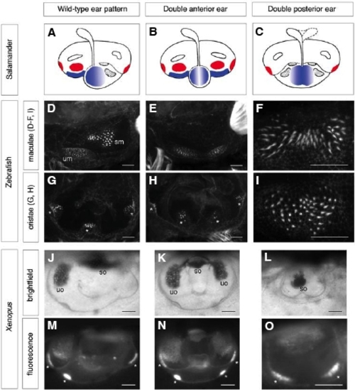
Examples of axial duplications in the ears of amphibian and zebrafish embryos. (A-C) Diagrams of ear phenotypes obtained by rotations of the otic rudiment about the AP axis in the salamander (reproduced, with permission, from Harrison, 1945). (A) Wild-type ear pattern; (B) double anterior ear; (C) double posterior ear. The utricular and saccular maculae (blue) and cristae (red) are highlighted for clarity. (D-I) Confocal images of ear phenotypes following manipulation of Hh signalling in the zebrafish embryo. Ears are stained with FITC-phalloidin to mark the hair bundles of sensory hair cells in the maculae (D-F, I) or cristae (G, H). (D,G) Wild-type pattern (86 hpf); (E,H) double anterior pattern (86 hpf) obtained by incubation of the embryo from the 10 somite stage to 22 hpf in 50 μM cyclopamine to inhibit Hh signalling; (F,I) two examples of a double posterior pattern (mirror-image saccular macula) obtained by injection of shha mRNA into the embryo at the 1-cell stage (72 hpf) (reproduced with permission of the Company of Biologists from Hammond et al., 2003). (J-O) Ear phenotypes at tadpole stage (stage 48) obtained by partial otic placode or otic vesicle ablations in the Xenopus embryo at stages 24-27 (reprinted with permission of Wiley-Liss, Inc., a subsidiary of John Wiley & Sons, Inc., from Waldman et al., 2007). (J,M) Wild-type pattern; (K,N) double anterior ear obtained by ablation of the posterior half of the otic placode; (L,O) double posterior ear obtained by ablation of the anterior half of the otic placode. In all double anterior ears, the utricular macula is duplicated, and four cristae are present. In the double posterior ears, the utricular macula is missing, and the number of cristae is reduced. In all panels, the anterior of the embryo is to the left; (A-I) are lateral views, (J-O) are dorsal views. Abbreviations: sm, saccular macula; so, saccular otolith; um, utricular macula; uo, utricular otolith. Asterisks indicate the position of cristae. Scale bars: (D-I) 25 μm; (J-O) 100 μm. |
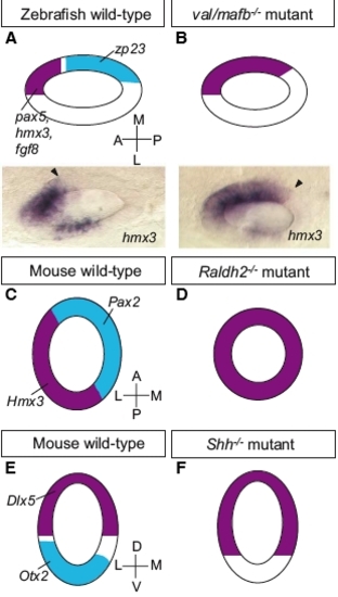
Examples of mutant phenotypes affecting axial patterning at the otic vesicle stage. (A,B) Disruption of the otic AP axis in the zebrafish val/mafb mutant (dorsal views). Expression of anterior markers ( hmx3, pax5, fgf8; purple) extends posteriorly around the medial wall of the otic vesicle, while expression of a posterior marker ( zp23; light blue) is lost (adapted from data in Kwak et al., 2002). The photographs show the expression of hmx3 at 25 hpf; arrowheads mark the posterior boundary of expression. (C,D) Disruption of the otic ML axis in the mouse Raldh2-/- mutant (dorsal views). Expression of a medial marker (Pax2; light blue) is lost, while expression of a lateral marker (Hmx3; purple) is expanded throughout the otic epithelium (adapted from data in Niederreither et al., 2000). (E,F) Disruption of the otic DV axis in the mouse Shh-/- mutant (transverse sections). Expression of the dorsal marker Dlx5 (purple) extends ventrally, while expression of the ventral marker Otx2 (light blue) is lost (adapted from data in Riccomagno et al., 2002). All drawings are schematic diagrams, not to scale; they show only a subset of the genes affected in each case. |