
|
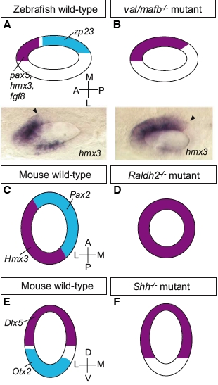
Fig. 3 Examples of mutant phenotypes affecting axial patterning at the otic vesicle stage. (A,B) Disruption of the otic AP axis in the zebrafish val/mafb mutant (dorsal views). Expression of anterior markers ( hmx3, pax5, fgf8; purple) extends posteriorly around the medial wall of the otic vesicle, while expression of a posterior marker ( zp23; light blue) is lost (adapted from data in Kwak et al., 2002). The photographs show the expression of hmx3 at 25 hpf; arrowheads mark the posterior boundary of expression. (C,D) Disruption of the otic ML axis in the mouse Raldh2-/- mutant (dorsal views). Expression of a medial marker (Pax2; light blue) is lost, while expression of a lateral marker (Hmx3; purple) is expanded throughout the otic epithelium (adapted from data in Niederreither et al., 2000). (E,F) Disruption of the otic DV axis in the mouse Shh-/- mutant (transverse sections). Expression of the dorsal marker Dlx5 (purple) extends ventrally, while expression of the ventral marker Otx2 (light blue) is lost (adapted from data in Riccomagno et al., 2002). All drawings are schematic diagrams, not to scale; they show only a subset of the genes affected in each case.