
- Title
-
Heart Regeneration in the Mexican Cavefish
- Authors
- Stockdale, W.T., Lemieux, M.E., Killen, A.C., Zhao, J., Hu, Z., Riepsaame, J., Hamilton, N., Kudoh, T., Riley, P.R., van Aerle, R., Yamamoto, Y., Mommersteeg, M.T.M.
- Source
- Full text @ Cell Rep.
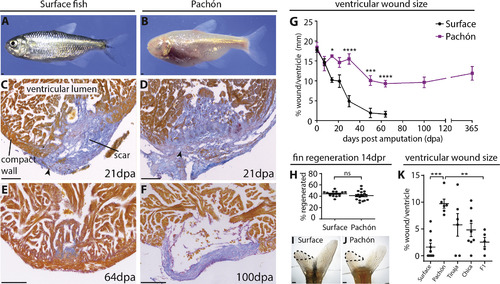
Permanent Scar Formation after Ventricular Resection in the Pachón Cavefish (A and B) Adult surface (A) and Pachón cavefish (B). (C–F) AFOG staining of the ventricular apex after resection. AFOG staining is a technique that stains myocardium orange and collagen blue. Both populations form a collagen scar (C and D, arrowheads), which disappears in the surface fish around 64 dpa (E), but persists in the Pachón (F). (G) Time line showing the reducing wound size in the surface fish but persisting wound in the Pachón. n ≥ 3 per population per time point, two-way ANOVA with Sidak’s test. (H) No difference in fin regeneration between Pachón (n = 18) and surface fish (n = 16) at 14 days post resection (dpr). Unpaired t test. (I and J) Regenerating dorsal lobes of tail fins of surface fish (I) and Pachón (J) at 14 dpr. Dotted line indicates the regenerated part. (K) Wound size 64 dpa in surface fish (n = 10), Pachón (n = 7), Tinaja (n = 6), Chica (n = 8), and surface fish × Pachón F1 hybrids (n = 5). One-way ANOVA with Tukey’s test. Detailed numbers and statistics are reported in STAR Methods Results are presented as mean ± SEM. All scale bars, 100 μm. |

lrrc10Is Required for Heart Regeneration in Zebrafish(A) RNAScope RNA expression analysis shows thatlrrc10is expressed specifically in all the MF20-positive myocardium at 7 dpa in surface fish and Pacho ́naswell as in zebrafish at 7dpi. Arrows point to absent expression in non-myocardial tissues, such as the wound, valves, and bulbus arteriosus. Expression aroundthe wound is higher in surface fish and zebrafish compared with Pacho ́n (arrowheads).(B)lrrc10is especially higher expressed in the compact myocardium (arrowheads) compared with the trabecular myocardium close to the wound (demarcatedwith line) in surface fish and zebrafish. This difference is absent in Pacho ́n.(C) Zebrafishlrrc10mutants were generated by removing 601 nucleotides, including the ATG start site using CRISPR/Cas9.(D) Genotyping with primers just outside the deleted region amplifies a product of 833 bp in wild-types and a product of 232 bp in mutants.(E) Whole-mountin situhybridization using anlrrc10antisense probe in 3 dpf (days post fertilization)lrrc10/(n = 12) and wild-type embryos (n = 25) shows thatlrrc10expression is specifically present in the wild-type heart (arrow) but absent in the mutant, confirming knockout oflrrc10.(F) AFOG staining showing large blue collagen scar in thelrrc10mutant compared with the wild-type control at 60 dpi.(G) Wound size at 60 dpi inlrrc10/(n = 7) and wild-type controls (n = 7), unpaired two-tailed t test.(H) PCNA/Mef2 staining onlrrc10/and controls at 7dpi.(I) No significant difference in the number of PCNA/Mef2-positive cells surrounding the wound inlrrc10/(n = 5) compared with wild-type hearts (n = 6) at 7 dpi.Unpaired two-tailed t test, equal variance.(J) PCNA/Mef2 staining onlrrc10/and controls at 7dpi at the base of the ventricle.(K) Increased number of PCNA/Mef2-positive cells on the other side of the ventricle inlrrc10/(n = 5) compared with wild-type hearts (n = 6) at 7 dpi. Unpairedtwo-tailed t test, equal variance.All scale bars, 100mm. BA, bulbus arteriosus; CW, compact wall; Lu, lumen; Va, valves. Results are presented as mean±SEM. EXPRESSION / LABELING:
PHENOTYPE:
|
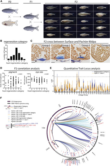
Three Loci Associated with Heart Regeneration Identified by QTL Analysis (A) The second generation of a cross between surface fish and Pachón results in offspring ranging from large, pigmented fish with no eyes to small cavefish-like fish with black eyes. One hundred eighty-eight F2 hearts were isolated 90 days after resection. (B) Number of F2 generation hearts per heart regeneration class. (C) AFOG staining on F2 hearts. Examples of four of the seven groups used for correlation tests and QTL analysis. Group 0, clear scar, neither compact wall myocardial thickening nor overgrowth (arrowhead). Group 2, clear scar, compact myocardium has started to grow over the scar (arrowhead). Group 4, clear scar, compact myocardium closed. Group 6, no sign of injury left. (D) No correlation between body melanocyte number (r = 0.085) or eye size (r = 0.089) and heart regeneration. Pearson’s correlation. (E) LOD scores per LG, showing three loci above the genome-wide significance threshold of 2.82 (95th percentile of maximum LOD from permutation analysis). (F) Circos plot mapping markers within LOD score peaks on LG1, LG9, and LG10 to the cavefish genome contigs and showing significant log2 fold expression changes for genes overlapping those loci. Detailed numbers and statistics are provided in STAR Methods. All scale bars, 100 μm. |