FIGURE SUMMARY
Title

Developmental temperature has persistent, sexually dimorphic effects on zebrafish cardiac anatomy

Authors
Dimitriadi, A., Beis, D., Arvanitidis, C., Adriaens, D., Koumoundouros, G.
Source
Full text @ Sci. Rep.

Multiple views from a single scan of an adult zebrafish. (A) Representative oblique slice defined by three landmarks (anterior and posterior end of the bulbus arteriosus, the centre of the 1st vertebra). (B) Inset of Fig. 2A, showing the ventricle (ven), bulbus arteriosus (ba), as well as the distance measurements taken. (C) Three-dimensional volume rendering of the bulbus arteriosus (purple) and ventricle (green). V, first vertebra. ba, bulbus arteriosus. ven, ventricle. Landmark 1, bulbus junction with the first branchial arch. Landmark 2, ventricle – bulbus valve. Landmark 3, ventricle apex. Landmarks 4 and 5 define the widest distance of the ventricle, perpendicularly to ventricle length (D2–3). D1–2, Bulbus-arteriosus length (BaL); D2–3, Ventricle length (VL); D4–5, maximum ventricle depth (VD), perpendicular to VL.

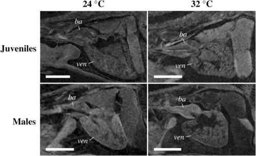
Representative primary images showing the comparatively more round ventricle in juvenile and male zebrafish reared at 32 °C developmental temperature (TD). Scale bars equal to 0.25 (juveniles) or 1.0 mm (males). ba, bulbus arteriosus. ven, ventricle.

Acknowledgments
This image is the copyrighted work of the attributed author or publisher, and ZFIN has permission only to display this image to its users. Additional permissions should be obtained from the applicable author or publisher of the image. Full text @ Sci. Rep.