
- Title
-
Zebrafish sp7 mutants show tooth cycling independent of attachment, eruption and poor differentiation of teeth
- Authors
- Kague, E., Witten, P.E., Soenens, M., Campos, C.L., Lubiana, T., Fisher, S., Hammond, C., Brown, K.R., Passos-Bueno, M.R., Huysseune, A.
- Source
- Full text @ Dev. Biol.
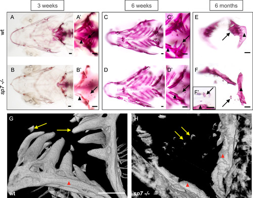
Defective tooth formation and lack of attachment insp7mutants observed throughout life. Alizarin Red S staining to show mineralization of the zebrafish pharyngeal teeth and fifth ceratobranchial (pharyngeal jaw) (A-F). A, A′, B, B′) In comparison to the wt, the fifth ceratobranchial (arrowheads) is poorly mineralized in mutants at 3 weeks post-fertilization, with only the extreme tip of the teeth stained (arrows). C, C′, D, D′) At 6 weeks, mineralization of the fifth ceratobranchial has increased in the mutants, but teeth remain as before. E, F, F′) sp7 mutant pharyngeal bone displays a more narrow and thinner morphology, and teeth do not have a mineralized attachment to the bone. G, H) Volume rendering pictures of micro-computed tomography (µCT) of zebrafish (>1.5 year) to show pharyngeal bones (arrowheads) and teeth (arrows) in wt (G) and sp7 mutant (H). Note the shape of the pharyngeal bones in the sp7 mutant and the absence of any bone of attachment. Scale bars represent 100 µm. PHENOTYPE:
|
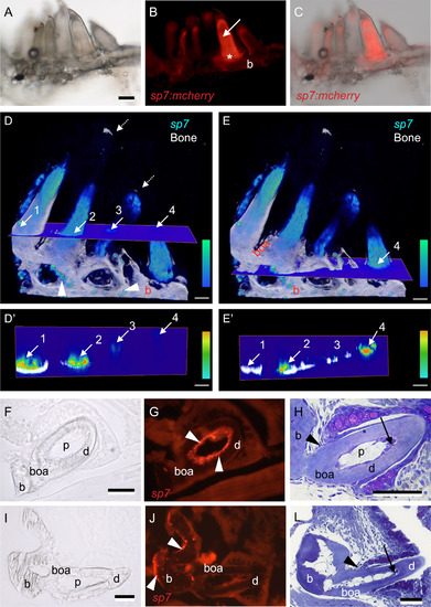
sp7expression in the pharyngeal bones and teeth of zebrafish. Whole mount (A-C), 3D renders from confocal images (D-E′) and immunostained frozen sections (F-G, I-J) of teeth in juvenile Tg(sp7:mcherry) zebrafish (14 mm SL), compared with toluidine blue stained sections of similar-sized wt zebrafish teeth (H, L). A) Juvenile zebrafish pharyngeal bone seen under transmitted light, with the full complement of teeth present. B) Same sample shown under fluorescent light revealing sp7 expression in the pulp (arrow), bone of attachment (asterisk) and underlying ceratobranchial bone (b). Note that other teeth show partial to no expression. C) Overlay between A and B; note that sp7 expression varies according to the maturation of each tooth. D-E) 3D volume renders of z-stack from confocal microscopy images of Tg(sp7:mcherry), level of sp7 expression is color-coded as indicated by a color bar (bottom right), bone is coloured in white. Note that expression is stronger in some teeth but partial in others. Four teeth are indicated with numbers; arrows indicate where a cross section hits each tooth in two different planes (D and E). Isolated osteoblasts expressing sp7 (arrowheads) are observed lining the pharyngeal bone (b), bone of attachment is clearly observed (boa). D′-E′) Corresponding cross section planes from D-E, one plane in a higher position (D′) than the other (E′). The same four teeth are indicated with arrows and numbers. Note that expression of sp7 is stronger in proximity of the mineralized dentin (tooth 1 and 2). F-G) Cross section of a young functional tooth, immunostained for sp7, seen in transmitted (F) and fluorescent (G) light. Note odontoblasts (white arrowheads) expressing sp7 in a linear arrangement juxtaposed to the dentin (d), continuing along the bone of attachment (boa). H) Toluidine blue stained plastic section of young functional tooth in a wt zebrafish. Note that odontoblasts at the tooth tip are still tall and polarized (black arrow). Black arrowhead indicates the level of the cervical loop and thus the limit between dentin (d) and bone of attachment (boa). I-J) Cross section of a mature functional tooth, immunostained for sp7, seen in transmitted (I) and fluorescent (J) light. Note lack of sp7 expression in the pulp (p), while osteoblasts (white arrowheads) express sp7 along the pharyngeal bone (b). L) Toluidine blue stained plastic section of a mature tooth in a wt zebrafish. Note that all odontoblasts have taken on a flattened shape, even in the tooth tip (black arrow). Black arrowhead indicates the level of the cervical loop and thus the limit between dentin (d) and bone of attachment (boa). Abbreviations: b, pharyngeal bone (fifth ceratobranchial); boa, bone of attachment; d, dentin; p, pulp. Scale bars represent 100 µm in (A-E′) and 50 µm in (F-L). EXPRESSION / LABELING:
|
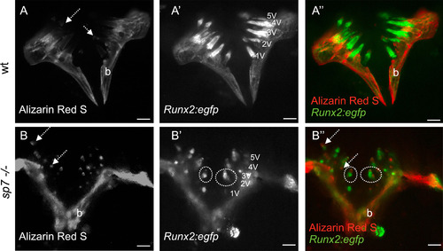
Tooth pattern insp7mutant is unaltered. A-B) wt and sp7 mutant carrying Tg(RUNX2:egfp) and live stained for Alizarin Red S followed by dissection of pharyngeal bone and observed under a stereomicroscope and fluorescent light. Alizarin Red S stains calcium deposition in mineralized tissues. Note tooth tips strongly stained in mutants (dashed arrows) (A,B). Despite the number of teeth in sp7 mutants being seemingly higher, it is still within the range that can be expected: each pharyngeal jaw can show as many as (maximum) 22 teeth (11 functional teeth, 11 replacement teeth). A′, B′) Tg(RUNX2:egfp) showing early odontoblasts and osteoblasts in pulp and bone respectively. Ventral tooth positions are indicated. Some tooth pulps display different shapes (dashed circles). Merged pictures with Alizarin Red S and RUNX2:egfp are shown (A″, B″). Scale bars represent 250 µm. PHENOTYPE:
|

Tooth abnormalities in thesp7mutant. Toluidine blue stained 2 µm sections of teeth in 6 months old wt (A, D and H) and sp7 mutant (B-C, E-F and J) zebrafish. Confocal imaging of teeth in 5 months wt and sp7 -/- zebrafish (G and I, respectively). A) Replacement tooth of a wt, with enameloid (arrowhead) and dentin (d) containing tubules (arrow), with predecessor tooth (asterisk) partly visible, localized in proximity of the pharyngeal epithelium. B) Overview of a defective tooth in the sp7 mutant with a thin layer of dentin (d) and highly cellular pulp (p). C) Magnification of a sp7 mutant tooth. Note small amount of dentin (d) and enameloid with traces of organic matter (arrowhead) but well organized enamel organ with polarized ameloblasts (asterisk). Note densely cellular pulp (p). D-E) Lower magnification picture to show the pharyngeal cavity (asterisks) and tooth arrangement in wt (D) and sp7 mutant (E). In wt, teeth are oriented with tooth tips (arrows) pointing to the pharyngeal cavity (asterisks), while in the sp7 mutant some teeth have an abnormal orientation with their tip (arrow) turned away from the pharyngeal cavity. Pairs of teeth can also be observed (arrowheads). F) A pair of tooth germs (predecessor, arrow and successor, arrowhead). These germs have an orientation relative to each other much as in the wt (compare with A). Note that the densely cellular pulps of both germs are interconnected without sharp boundary. G-J) Confocal imaging (G, I) and histological sections (H, J) of similar teeth to show pulps in wt (G, H) and sp7 mutant (I, J). G, I). wt (G) and sp7 mutant (I) carrying (Tg(RUNX2:egfp) and stained for Alizarin Red S, imaged using confocal microscopy followed by 3D projection. The pharyngeal cavity region (asterisks), pharyngeal bone (b) and bone of attachment (boa, in wt only) are labelled. Numbers label the different teeth. Note that the bone of attachment delimits each pulp (p) in the wt (G) while in sp7 mutants (I) the lack of bones of attachment leads to abnormally connected pulps (p). H, J) Toluidine blue stained 2 µm cross section of pulps (p) in wt (H) and multi-pulps (p) connected in the sp7 mutant (J). The pharyngeal cavity region is indicated by asterisks and the bone of attachment (boa) is labelled in the wt. Scale bars A, B, F and J = 50 µm; C = 20 µm; D, E, G, H and I= 100 µm. PHENOTYPE:
|

Osteoclastic activity revealed by TRAP staining. TRAP activity (red staining, arrows) in the predecessor tooth in wt (A) and sp7 mutant (C) zebrafish, likely elicited by growth of a replacement tooth (asterisk in C). Inset in C shows magnification of TRAP activity involved in removal of the dentin (d). Note small size of the mutant teeth compared to the wt (A). TRAP activity (red staining, arrows) involved in remodelling of the ceratobranchial bone opposite a developing tooth (asterisk) is not different between wt (B) and mutant (D) fish. Scale bars = 50 µm. PHENOTYPE:
|
Reprinted from Developmental Biology, 435(2), Kague, E., Witten, P.E., Soenens, M., Campos, C.L., Lubiana, T., Fisher, S., Hammond, C., Brown, K.R., Passos-Bueno, M.R., Huysseune, A., Zebrafish sp7 mutants show tooth cycling independent of attachment, eruption and poor differentiation of teeth, 176-184, Copyright (2018) with permission from Elsevier. Full text @ Dev. Biol.