
Fig. 2
- ID
- ZDB-FIG-180622-10
- Publication
- Kague et al., 2018 - Zebrafish sp7 mutants show tooth cycling independent of attachment, eruption and poor differentiation of teeth
- Other Figures
- All Figure Page
- Back to All Figure Page
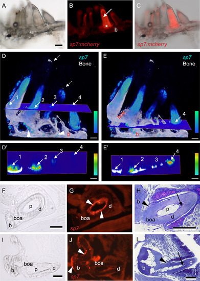
sp7expression in the pharyngeal bones and teeth of zebrafish. Whole mount (A-C), 3D renders from confocal images (D-E′) and immunostained frozen sections (F-G, I-J) of teeth in juvenile Tg(sp7:mcherry) zebrafish (14 mm SL), compared with toluidine blue stained sections of similar-sized wt zebrafish teeth (H, L). A) Juvenile zebrafish pharyngeal bone seen under transmitted light, with the full complement of teeth present. B) Same sample shown under fluorescent light revealing sp7 expression in the pulp (arrow), bone of attachment (asterisk) and underlying ceratobranchial bone (b). Note that other teeth show partial to no expression. C) Overlay between A and B; note that sp7 expression varies according to the maturation of each tooth. D-E) 3D volume renders of z-stack from confocal microscopy images of Tg(sp7:mcherry), level of sp7 expression is color-coded as indicated by a color bar (bottom right), bone is coloured in white. Note that expression is stronger in some teeth but partial in others. Four teeth are indicated with numbers; arrows indicate where a cross section hits each tooth in two different planes (D and E). Isolated osteoblasts expressing sp7 (arrowheads) are observed lining the pharyngeal bone (b), bone of attachment is clearly observed (boa). D′-E′) Corresponding cross section planes from D-E, one plane in a higher position (D′) than the other (E′). The same four teeth are indicated with arrows and numbers. Note that expression of sp7 is stronger in proximity of the mineralized dentin (tooth 1 and 2). F-G) Cross section of a young functional tooth, immunostained for sp7, seen in transmitted (F) and fluorescent (G) light. Note odontoblasts (white arrowheads) expressing sp7 in a linear arrangement juxtaposed to the dentin (d), continuing along the bone of attachment (boa). H) Toluidine blue stained plastic section of young functional tooth in a wt zebrafish. Note that odontoblasts at the tooth tip are still tall and polarized (black arrow). Black arrowhead indicates the level of the cervical loop and thus the limit between dentin (d) and bone of attachment (boa). I-J) Cross section of a mature functional tooth, immunostained for sp7, seen in transmitted (I) and fluorescent (J) light. Note lack of sp7 expression in the pulp (p), while osteoblasts (white arrowheads) express sp7 along the pharyngeal bone (b). L) Toluidine blue stained plastic section of a mature tooth in a wt zebrafish. Note that all odontoblasts have taken on a flattened shape, even in the tooth tip (black arrow). Black arrowhead indicates the level of the cervical loop and thus the limit between dentin (d) and bone of attachment (boa). Abbreviations: b, pharyngeal bone (fifth ceratobranchial); boa, bone of attachment; d, dentin; p, pulp. Scale bars represent 100 µm in (A-E′) and 50 µm in (F-L). |
| Gene: | |
|---|---|
| Fish: | |
| Anatomical Terms: | |
| Stage: | Adult |
Reprinted from Developmental Biology, 435(2), Kague, E., Witten, P.E., Soenens, M., Campos, C.L., Lubiana, T., Fisher, S., Hammond, C., Brown, K.R., Passos-Bueno, M.R., Huysseune, A., Zebrafish sp7 mutants show tooth cycling independent of attachment, eruption and poor differentiation of teeth, 176-184, Copyright (2018) with permission from Elsevier. Full text @ Dev. Biol.