
- Title
-
An optimized method for counting dopaminergic neurons in zebrafish
- Authors
- Matsui, H., Sugie, A.
- Source
- Full text @ PLoS One
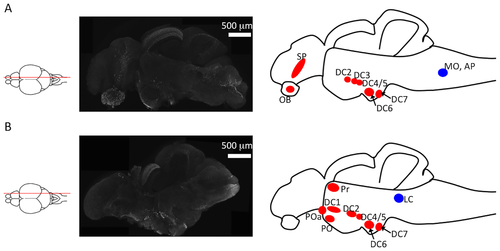
Identification of tyrosine hydroxylase (TH)+ neurons in sagittal sections of zebrafish brains. (A) Left panel: an illustration of the sectioning position; middle panel: TH+ immunoreactivity in a microsliced section of a 6-month-old zebrafish brain; right panel: TH+ neuron clusters found in this section. The red and blue circles indicate dopaminergic and noradrenergic neurons, respectively. (B) Left panel: an illustration indicating the sectioning position. The middle panel is an immunostaining of TH using a microsliced section of 6-month-old zebrafish. The right panel illustrates the TH+ neuron clusters in this section. The red and blue circles indicate dopaminergic and noradrenergic neurons, respectively. DC1?7: diencephalic catecholaminergic cluster. Abbreviations: AP, area postrema; LC, locus coeruleus; MO, medulla oblongata interfascicular zone and vagal area; OB, olfactory bulb; PO, preoptic region; POa, anterior preoptic region; Pr, dorsal pretectum; SP, subpallium. EXPRESSION / LABELING:
|

Tyrosine hydroxylase (TH)+ neurons in the axial sections of zebrafish brains. (A) The illustration indicates the position of sectioning in panels B?E. (B?E) Immunostaining of TH using a microsliced section of 6-month-old zebrafish. Different rostro?caudal levels show the localization of TH+ dopaminergic neurons in the olfactory bulb and subpallium (B); pretectum, posterior periventricular preoptic nucleus, and suprachiasmatic nucleus (C); pretectum, posterior tuberculum, and paraventricular organ (D); and posterior recess of the diencephalic ventricle (E). TH+ noradrenergic neurons are distributed in the locus coeruleus (F). (G) The number of TH+ neurons in the posterior tuberculum (dopaminergic neurons) and the locus coeruleus (noradrenergic neurons). The bars represent SEM (n = 10). (D?, F?) TH immunoreactivity in a paraffin section of a 6-month-old zebrafish. (H) The counted number of TH+ neurons using paraffin sections in the posterior tuberculum (dopaminergic neurons) and the locus coeruleus (noradrenergic neurons). Bars represent the SEM (n = 6). EXPRESSION / LABELING:
|
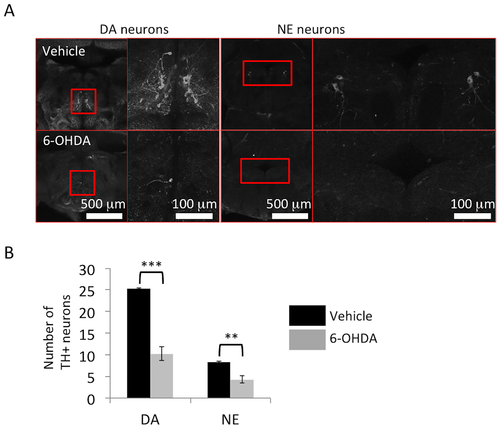
Reduction of tyrosine hydroxylase (TH)+ neurons after 6-hydroxydopamine (6-OHDA) administration. (A) Representative images of dopaminergic neurons in the posterior tuberculum and noradrenergic neurons in the locus coeruleus. Right panel: magnified pictures of the red square regions are shown. (B) The numbers of dopaminergic neurons in the posterior tuberculum and noradrenergic neurons in the locus coeruleus are significantly decreased by 6-OHDA treatment in 12-month-old zebrafish; n = 5 fish per group. Bars represent the SEM. **, p < 0.01, ***, p < 0.001. |
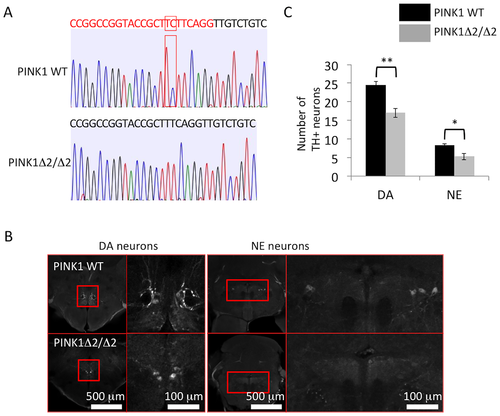
Reduction of tyrosine hydroxylase (TH)+ neurons in PINK1-deficient (PINK1-KO) zebrafish. (A) Sequence information of PINK1-deficient zebrafish (c.176_177del, PINK1?2/?2). The red letter indicates the target sequence of the guide RNA, and the red square indicates the site of the 2-bp deletion. (B) Representative images of dopaminergic neurons in the posterior tuberculum and noradrenergic neurons in the locus coeruleus. Right panel: magnified pictures of the red square regions. (C) The numbers of dopaminergic neurons in the posterior tuberculum and noradrenergic neurons in the locus coeruleus were significantly decreased in PINK1-deficient zebrafish (PINK1?2/?2) (4-month-old zebrafish); n = 5 fish per group. Bars represent the SEM. *, p < 0.05, **, p < 0.01. |