
FIGURE
Fig. 3
- ID
- ZDB-FIG-171006-4
- Publication
- Matsui et al., 2017 - An optimized method for counting dopaminergic neurons in zebrafish
- Other Figures
- All Figure Page
- Back to All Figure Page
Fig. 3
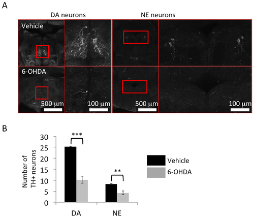
Reduction of tyrosine hydroxylase (TH)+ neurons after 6-hydroxydopamine (6-OHDA) administration. (A) Representative images of dopaminergic neurons in the posterior tuberculum and noradrenergic neurons in the locus coeruleus. Right panel: magnified pictures of the red square regions are shown. (B) The numbers of dopaminergic neurons in the posterior tuberculum and noradrenergic neurons in the locus coeruleus are significantly decreased by 6-OHDA treatment in 12-month-old zebrafish; n = 5 fish per group. Bars represent the SEM. **, p < 0.01, ***, p < 0.001. |
Expression Data
| Antibody: | |
|---|---|
| Fish: | |
| Condition: | |
| Anatomical Terms: | |
| Stage: | Adult |
Expression Detail
Antibody Labeling
Phenotype Data
| Fish: | |
|---|---|
| Condition: | |
| Observed In: | |
| Stage: | Adult |
Phenotype Detail
Acknowledgments
This image is the copyrighted work of the attributed author or publisher, and
ZFIN has permission only to display this image to its users.
Additional permissions should be obtained from the applicable author or publisher of the image.
Full text @ PLoS One