
- Title
-
Improved Long-Term Imaging of Embryos with Genetically Encoded ?-Bungarotoxin
- Authors
- Swinburne, I.A., Mosaliganti, K.R., Green, A.A., Megason, S.G.
- Source
- Full text @ PLoS One

Tricaine and isoeugenol co-operate towards healthier immobilization. (A) Heat map of percent immobile for 48 combinations of tricaine (0?200 �g/ml) and isoeugenol (0?0.003% v/v). Embryos were dechorionated and soaked from 24?27 hpf when they were assayed for immobility. (B) Continuation of treatments from (A), embryos were assayed for immobility at 72 hpf. (C,D) Representative micrographs of control (C) and 200 �g/ml tricaine treated (D) embryos. Arrow in (D) shows failure of semicircular canal projection fusion. Asterisk in (D) shows pericardial edema. (E) Heat map of percent of embryos with pericardial edema at 72 hpf. (F) Percent control otic vesicle diameter (OVD) was calculated by dividing the average of 10?30 experimental embryos by the average of 10?30 control embryos. Heat map of percent control OVD for the combinatorial treatments. OVD was measured at 72 hpf using micrographs like those in (C, D). Percentage is based on normalization to untreated control. (G) Merge of heatmaps from (A, 27 hpf) and (F) that highlights the tradeoffs between embryo immobility and healthy development. |
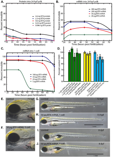
α-bungarotoxin immobilizes embryos while permitting normal development. (A) Percent of embryos immobile after injection of α-bungarotoxin protein (0.046?4.6ng) into the yolk at 24 hpf. (B) Percent of embryos immobile after injection of α-bungarotoxin mRNA (20?400 pg) into the yolk at 24 hpf. (C) Percent of embryos immobile after injection of of α-bungarotoxin mRNA (5?100 pg) into the 1-cell zygote. (D) Percent control OVD at 72 hpf for injection of α-bungarotoxin mRNA into the 1-cell zygote (green), into the yolk (yellow), and reference anesthetic treatments that permitted long-term immobilization (blue). (*) Not significantly different from control, Mann-Whitney-Wilcoxon two tailed P-value 0.87. (?) Significantly different from control, Mann-Whitney-Wilcoxon two tailed P-value 0.0011. (E, G) Control embryo at 72 hpf that was injected with 50 pg of membrane-citrine mRNA into the 1-cell zygote. (F, H) 72 hpf embryo that was injected with 50 pg of α-bungarotoxin mRNA into the 1-cell zygote. (I) Control larva at 8 days post fertilization (dpf) injected with 50 pg of membrane-citrine mRNA into the 1-cell zygote. (J) 8 dpf larva that was injected with 50 pg of α-bungarotoxin mRNA into the 1-cell zygote. |
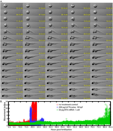
Long-term imaging of embryos immobilized with α-bungarotoxin mRNA. (A) Montage of an immobilized embryo?s development from the 1-cell stage to 85 hpf after it had been injected with 50 pg of α-bungarotoxin mRNA into the 1-cell. Images are shown from every hour of development. (B) Quantification of the full time-course that included 153,452 images that were acquired every 2 seconds. The movement index was calculated as the maximum difference between each image and its subsequent image in the time-series. The index was normalized to the average maximum difference in the first 2,000 time points. Control embryos (red) and embryos in 200 �g/ml tricaine (blue) begin twitching at around 18 hpf and then may swim out of the field while α-bungarotoxin injected embryos (green) showed very little movement until 80 hpf. |
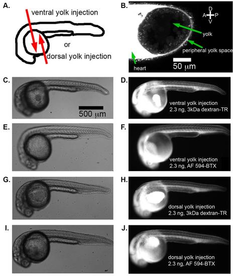
α-bungarotoxin protein injected into the yolk is distributed throughout the embryo. (A) Two injection strategies explored for α-bungarotoxin protein injection: either into the ventral side of the yolk or the dorsal side of the yolk of zebrafish embryos at 24 hpf. (B) Fluorescence from 2.3 ng Alexa-Fluor 594 conjugated α-bungarotoxin injected into the ventral yolk imaged by laser-scanning confocal microscopy. The peripheral yolk space appears continuous with the fluid entering the heart. DIC images (left) and fluorescent images (right) of representative embryos receiving ventral yolk injections of 2.3 ng 3 kDa dextran-Texas red (C-D), ventral yolk injections of 2.3 ng Alexa-Fluor 594 conjugated α-bungarotoxin (E-F), dorsal yolk injections of 2.3 ng 3 kDa dextran-Texas red (G-H), and dorsal yolk injections of 2.3 ng Alexa-Fluor 594 conjugated α-bungarotoxin (I-J). Scale bar in (C) applies for (C-J). |