
- Title
-
Phenotype classification of zebrafish embryos by supervised learning
- Authors
- Jeanray, N., Marée, R., Pruvot, B., Stern, O., Geurts, P., Wehenkel, L., Muller, M.
- Source
- Full text @ PLoS One
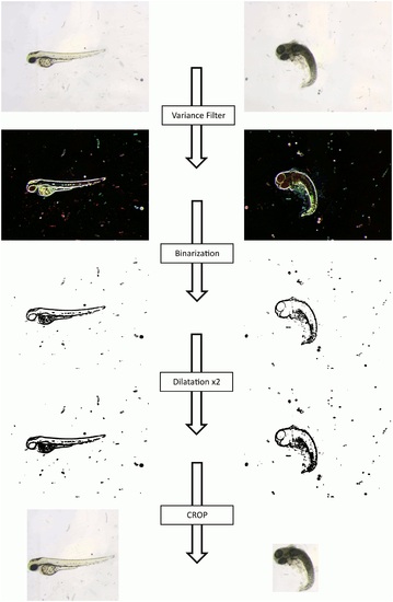
Image Preprocessing: From original image to the one used for classification. The original image is submitted to an ImageJ script (S1 Fig. for the code) which allows to automatically crop the zebrafish embryo into a square as far as possible. The embryo will be surrounded by a rectangle if it is placed near the sides of the original image. We use a connected-component labeling approach combined with different morphological transformations and binarizations, in order to localize the embryo. |

Examples of images representing all the analyzed phenotypes. “Without phenotype” defect, also called “Normal” embryos (A) are without any phenotype. The “Dead” phenotype (B) is shown as totally necrosed, while the “Chorion” phenotype (C) represents embryos that are still located in their chorion. In “Down Curved Tail” (D), the tail is obviously oriented downward compared to the horizontal. “Hemostasis” (E) presents a small amount of blood which can be located everywhere in the embryo (mainly in the head or in the pericardial area). “Necrosed Yolk Sac” (F) corresponds to a darker yolk compared to normal. In the “Edema” phenotype (G), an edema generally surrounds the anteroventral part of the fish. The “Short Tail” phenotype (H) describes a tail shorter than normal. “Up Curved Fish” (I) and “Up Curved Tail” (J) are two slightly different phenotypes. There is a curvature on the back of the embryo for “Up Curved Fish” (like a kind of lordosis), whereas the curvature is located on the tail for the “Up Curved Tail” phenotype. |

Examples of images annotated as “Chorion” and “Dead” phenotypes misclassified by the two-tier approach. This Fig. shows the confusion made by the two-tiered approach between “Chorion” and “Dead” classes for some sensitive images. In column “A”, three images are annotated as belonging to the “Dead” class and classified by the two-tiered approach as “Chorion” because of their rounded shape and in some cases, the not totally necrosed embryo. On the other hand, column “B” shows examples of images annotated as “Chorion” but classified as “Dead” because of the necrosed aspect of the embryos. |

Examples of sensitive images for “Necrosed Yolk Sac” and “Up Curved Tail” phenotypes. This Fig. shows examples of images where the impact of the subjectivity of the experts during image annotation could explain the weak classification results for the two phenotypes “Necrosed Yolk Sac” and “Up Curved Tail”. Columns “A” and “B” show examples of images annotated as belonging, respectively to the “Necrosed Yolk Sac” and the “no_Necrosed Yolk Sac” classes. These images are actually very similar even if they were annotated as belonging to opposite classes. The same observation is made for images in columns “C” and “D”. Images in “C” are annotated by experts as belonging to the “Up Curved Tail” class whereas images in “D” are annotated as not belonging to the “Up Curved Tail” class. |