
- Title
-
Apical localization of ASIP/PAR-3:EGFP in zebrafish neuroepithelial cells involves the oligomerization domain CR1, the PDZ domains, and the C-terminal portion of the protein
- Authors
- von Trotha, J.W., Campos-Ortega, J.A., and Reugels, A.M.
- Source
- Full text @ Dev. Dyn.
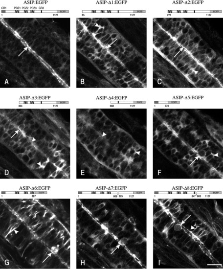
The PDZ domains are necessary for the localization of ASIP/PAR-3:EGFP to the apical cortex. A-C: Upon deletion of a single PDZ domain, the corresponding fusion proteins still localize at the apical cortex in essentially the same way as the full-length ASIP/PAR-3:EGFP protein. D: The apical localization is severely impaired upon deletion of all three PDZ domains, although a considerable portion of the fusion protein still localizes to the apical cortex. Apical membrane localization is denoted by arrows. A schematic representation of the different constructs is depicted above the confocal image. Anterior is toward the upper left. Scale bar = 20 μm. |
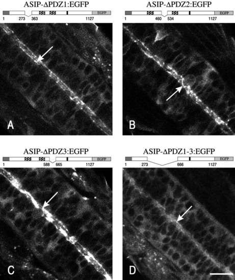
The CR1 oligomerization domain, the PDZ domains, and the C-terminal part are involved in apical membrane localization of ASIP/PAR-3:EGFP. A-I: Localization of the various ASIP/PAR-3:EGFP fusion proteins. See text for details. Apical membrane localization is denoted by arrows. B-F: Arrowheads mark localization in the cytoplasm. G,I: Arrowheads point to a localization at the lateral cortex. Note: deletion of the 15 amino acids (aa) in the C-terminal part of ASIP/PAR-3 yields the Pard3b isoform. Anterior is toward the upper left. Scale bar = 20 μm. |
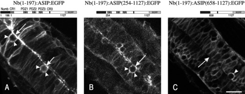
The apical localization signals in ASIP/PAR-3 prevent the basolateral localization of a Numb:PAR-3 fusion protein. A-C: Localization of the different EGFP-tagged Numb:ASIP fusion constructs. See text for details. A,B: Apical membrane localization is denoted by arrows, whereas arrowheads point to a localization at the basolateral cell cortex. Note: Decreased resolution in B is due to a shorter confocal scanning time. C: Numb(1-196):ASIP(658-1127):EGFP is mainly localized to the basolateral membrane (arrowhead), resulting in an apical cortex that is almost completely devoid of the fusion protein (arrow). Anterior is toward the upper left. Scale bar = 20 μm. |