
- Title
-
Molecular characterization of calymmin, a novel notochord sheath-associated extracellular matrix protein in the zebrafish embryo
- Authors
- Cerdà, J., Gründ, C., Franke, W.W., and Brand, M.
- Source
- Full text @ Dev. Dyn.

Expression of Calymmin mRNA during zebrafish embryogenesis as detected by whole-mount in situ hybridization (A-F) and Northern blot (G). Lateral views of embryos with rostral to the left (A,B,D-F) and 2- to 3-µm cross-section (C). A-C: Expression of the mRNA in notochord precursor cells in 15-somite stage embryos; C shows that transcripts are specifically expressed by the vacuolated notochord cells. D,E: Pharyngula period (24 hours postfertilization [hpf]). In D, the expression of c29-77 cDNA in the notochord shows a gradient with a higher level of transcripts toward the cells located in the tail region. E shows specific expression in the notochord cells at the level of the trunk, whereas both the adjacent floor plate (asterisk) and hypochord (arrowhead) cell layers are negative; the notochord sheath is indicated by an arrow. F: 34 hpf-embryos showing that expression becomes restricted to notochord cells located in the posterior region of the tail (F), where the notochord cells are differentiating. The results shown were obtained by using DIG-labelled riboprobes derived from full-length c29-77 (3.273 kb) and are identical when using riboprobes synthesized from c39a (4.049 kb). G: Northern blot showing the detection of a ~4.1-kb mRNA in 14- to 19-somite-stage zebrafish embryos (lane 1), whereas the message was not detected in adult fish (lane 2). Thirty micrograms of total RNA were loaded per lane. The size of molecular markers and ribosomal RNA is indicated on the left. EXPRESSION / LABELING:
|
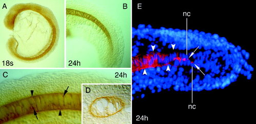
Localization of Calymmin in wild-type zebrafish embryos by whole-mount immunocytochemistry (A-D) and immunofluorescence microscopy (E). Views of embryos with rostral to the left (A-C), cross-section of labelled embryos (D), and cryostat section (E). A: 18-somite stage embryo showing staining within the developing notochord. B,C: Embryos at the pharyngula period (24 hours postfertilization [hpf]) showing Calymmin staining at the notochord sheath (C), which is less intense at the level of the tail (B). The arrowheads point to the notochord sheath, whereas the arrows indicate subcellular staining deposits close to the nucleus of the notochord cells. D: Cross-section of labelled embryos at the level of the trunk showing positive and specific immunolocalization of Calymmin in the notochord sheath. Note the high vacuolization of the notochord cells that are surrounded by the sheath. E: Immunofluorescence localization of Calymmin (red color) in the notochord cells located in the tail and anterior trunk of 24-hpf embryos. The nucleus of the notochord cells can be visualized by the Hoechst fluorescent dye (blue color). The arrows indicate the juxtanuclear localization of Calymmin within the notochord cells, likely in the Golgi apparatus, whereas the arrowheads point to the appearance of Calymmin at the notochordal sheath. nc, nucleus of the notochord cells. EXPRESSION / LABELING:
|

Detection of Calymmin in wild-type and ntl mutant zebrafish at 22 hours postfertilization embryos. A,B: General views of wild-type (A) and ntl mutant (B) embryos (rostral to the left) after whole-mount immunocytochemistry. Note the absence of the notochord and tail in the ntl mutants, in which Calymmin was not detected. C: Coomassie brilliant blue-stained sodium dodecyl sulphate-polyacrylamide gel electrophoresis of cytoskeletal fractions from wild-type (lane 1) and ntl embryos (lane 2), and the corresponding immunoblot (lanes 1′ and 2′), showing the absence of the anti-Calymmin reactive band in the mutant embryos. The relative molecular masses of the reference proteins are indicated on the left. EXPRESSION / LABELING:
PHENOTYPE:
|
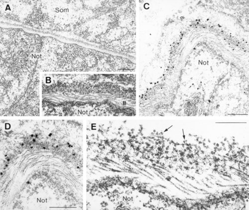
Ultrastructure of the notochordal sheath in 24-hours postfertilization zebrafish embryos (A,B,E), and ultrastructural localization of Calymmin (C,D). A: General view of two adjacent notochord cells surrounded by the notochord sheath, separating the notochord from the developing somites (Som). Note the presence of vacuoles and secretory vesicles within the notochord cells. B: Electron photomicrograph of the notochordal sheath, showing a proximal (to the notochord cells) basal lamina (I), a perinotochordal region of clearly striated fibrils (II), and an outer loosely organized and granular matrix (III). C,D: Immunogold localization of Calymmin at the outer granular layer of the notochordal sheath, whereas the rest of the sheath compartments appears unlabelled by the antibody. E: Ultrastructure of the notochordal sheath after conventional electron microscopy procedures in the presence of high concentration of detergents to unravel its organization. The granular-attached fibrillar nature of the Calymmin-labelled sheath compartment is indicated by arrows. Below it lies the microfibrillar layer, showing a characteristic collagen-like cross-banding pattern, and the basal lamina of the notochord cells. Not, notochord cells. Scale bars = 2.5 µm in A, 0.25 µm in B, 1 µm in C, 0.5 µm in D, 0.3 µm in E. EXPRESSION / LABELING:
|