
Fig. 6
- ID
- ZDB-FIG-130228-3
- Publication
- Cerdà et al., 2002 - Molecular characterization of calymmin, a novel notochord sheath-associated extracellular matrix protein in the zebrafish embryo
- Other Figures
- All Figure Page
- Back to All Figure Page
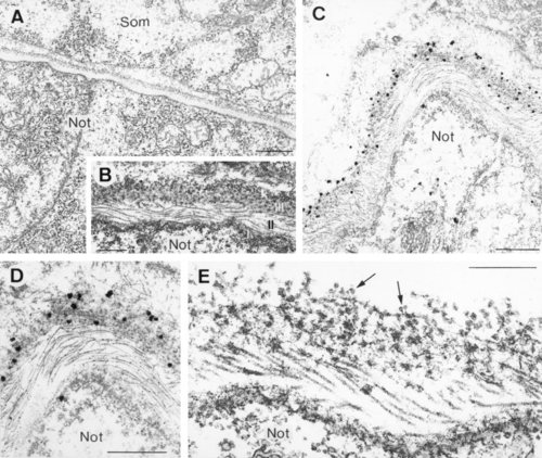
Ultrastructure of the notochordal sheath in 24-hours postfertilization zebrafish embryos (A,B,E), and ultrastructural localization of Calymmin (C,D). A: General view of two adjacent notochord cells surrounded by the notochord sheath, separating the notochord from the developing somites (Som). Note the presence of vacuoles and secretory vesicles within the notochord cells. B: Electron photomicrograph of the notochordal sheath, showing a proximal (to the notochord cells) basal lamina (I), a perinotochordal region of clearly striated fibrils (II), and an outer loosely organized and granular matrix (III). C,D: Immunogold localization of Calymmin at the outer granular layer of the notochordal sheath, whereas the rest of the sheath compartments appears unlabelled by the antibody. E: Ultrastructure of the notochordal sheath after conventional electron microscopy procedures in the presence of high concentration of detergents to unravel its organization. The granular-attached fibrillar nature of the Calymmin-labelled sheath compartment is indicated by arrows. Below it lies the microfibrillar layer, showing a characteristic collagen-like cross-banding pattern, and the basal lamina of the notochord cells. Not, notochord cells. Scale bars = 2.5 µm in A, 0.25 µm in B, 1 µm in C, 0.5 µm in D, 0.3 µm in E. |
| Gene: | |
|---|---|
| Antibody: | |
| Fish: | |
| Anatomical Term: | |
| Stage: | Prim-5 |