
|
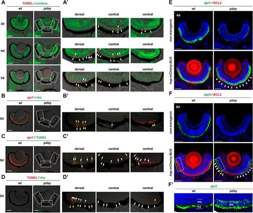
Fig. 3 Rods and cones undergo apoptosis in pday mutants. (A) TUNEL (red) of wild-type and pday mutant retinas. Nuclei were counterstained with Sytox Green (green). (A′) Higher magnification of the dorsal, central and ventral ONL of pday mutant retinas, which are indicated by white rectangles in A. White arrowheads indicate TUNEL signals (red). (B) Double labeling of 5-dpf wild-type and pday mutant retinas with zpr1 (red) and anti-rhodopsin (green) antibodies. (B′) Higher magnification of the dorsal, central and ventral ONL of pday mutant retinas, which are indicated by white rectangles in B. Both rhodopsin (yellow arrowheads) and zpr1 signals were observed in the peripheral region of the dorsal and ventral ONL. In addition, zpr1-positive flattened cells were located in the central ONL (white arrow). (C) TUNEL (green) of 5-dpf wild-type and pday mutant retinas combined with zpr1 antibody labeling (red). (C′) Higher magnification of the dorsal, central and ventral ONL of pday mutant retinas, indicated by white rectangles in C. Many TUNEL signals were associated with zpr1 signals (white arrows); however, a few were not associated with zpr1 (white arrowheads). (D) TUNEL (red) of 5-dpf wild-type and pday mutant retinas combined with anti-rhodopsin antibody labeling (green). (D′) Higher magnification of the dorsal, central and ventral ONL of pday mutant retinas, indicated by white rectangles in D. Rhodopsin signals were detected in the peripheral region of dorsal and ventral ONL (yellow arrowheads). TUNEL signals associated with rhodopsin signals were observed only in the peripheral region of the dorsal and ventral ONL (white arrowheads). TUNEL signals were not associated with rhodopsin signals in the central ONL and non-peripheral regions of the dorsal and ventral ONL (white arrows). (E,F) Wild-type and pday mutant retinas with and without the transgene Tg[hsp: mCherry-Bcl2], labelled with zpr1 (E) and zpr3 (F) antibodies. Nuclei were counterstained with Hoechst 33342 (blue). Both zpr1 and zpr3 signals in the ONL were recovered in pday mutant retinas with Tg[hsp: mCherry-Bcl2] (white arrowheads). (F′) Higher magnification of the dorsal ONL indicated by white rectangles in F. Sample sizes are shown in Table S3. Scale bars: 50 µm (A-D); 40 µm (E,F).